At Craft Body Scan, our board-certified radiologists review thousands of abdominal studies each year. That volume surfaces a pattern most patients never hear from their doctors – a pattern that begins with a normal test result and a pain that refuses to go away.
You went to the doctor for persistent upper-right abdominal pain. The doctor ordered a gallbladder ultrasound. The results came back unremarkable.
And the pain is still there.
That scenario plays out constantly. A gallbladder ultrasound is fast, affordable, and excellent at detecting gallstones – but it carries real blind spots. When symptoms point toward a complication or a problem in an adjacent organ, ultrasound alone often fails to deliver the full picture.
Understanding what this first-line scan can and cannot detect is the step that separates patients who get answers from patients who keep cycling through inconclusive appointments. A gallbladder ultrasound uses sound waves to produce real-time images of the gallbladder, bile ducts, and surrounding structures.
For simple gallstone cases, it performs well. The moment disease escalates beyond basic stone presence – toward gangrenous cholecystitis, perforation, abscess, or simultaneous pancreatic involvement – ultrasound begins to miss what matters most.
That is when a CT scan for gallbladder disease, a HIDA scan, or an MRI becomes the right tool. This article breaks down exactly what a gallbladder ultrasound shows, where it falls short, and how to identify when advanced imaging is the next step you actually need.
What a Gallbladder Ultrasound Actually Shows

A gallbladder ultrasound is the standard first-line study for right upper quadrant pain – and for good reason. It takes under 30 minutes, uses no ionizing radiation, requires no contrast agent, and is available at virtually every imaging center in the country. Radiologists use it to evaluate the gallbladder and biliary system with reasonable accuracy for the most common conditions.
To understand how a gallstone imaging study works in practice, it helps to know what ultrasound is physically capable of detecting. The table below outlines what this modality reliably shows versus what it frequently misses.
| Finding | Ultrasound Performance | Clinical Significance |
|---|---|---|
| Gallstones | High sensitivity (94-98%) | Detects stones as small as 2-3mm in a properly distended gallbladder |
| Gallbladder wall thickening | Moderate accuracy | Suggests acute cholecystitis; finding is nonspecific and also seen in liver disease and heart failure |
| Pericholecystic fluid | Moderate accuracy | Indicates active inflammation; helps support acute cholecystitis diagnosis alongside other findings |
| Common bile duct dilation | Good sensitivity; limited in patients with excess bowel gas or obesity | Suggests obstruction from a migrated stone or mass |
| Gangrenous cholecystitis | Poor sensitivity (20-50%) | A life-threatening complication frequently missed on ultrasound alone |
| Gallbladder perforation or abscess | Very limited | Requires CT confirmation to identify extent of perforation and infection |
| Pancreatic involvement | Limited due to overlying bowel gas | Gallstone pancreatitis from stone migration rarely visible on ultrasound |
| Gallbladder function and ejection fraction | Not measurable | Requires HIDA scan for any assessment of gallbladder contractile function |
The pattern is clear. For stone detection, ultrasound is a reliable tool. For complications, it struggles – and those complications are exactly what land patients in the emergency department or the operating room.
This gap between what ultrasound can and cannot see is not a flaw of the technology. It is simply the nature of what sound waves do when they encounter complex pathology, fat tissue, or bowel gas. Knowing these limits is what allows patients and physicians to make informed decisions about when a gallbladder ultrasound is sufficient and when it is not the right tool for the job.
Why a Normal Gallbladder Ultrasound Does Not Always Mean Normal
This is one of the most consequential misunderstandings in abdominal imaging. A normal gallbladder ultrasound does not rule out cholecystitis, biliary dyskinesia, early gallbladder cancer, or a gallstone that has migrated into the bile duct.
What it rules out is visible stones inside a gallbladder that was adequately visualized. Those are meaningfully different things.
Acalculous cholecystitis – gallbladder inflammation without stones – affects roughly 5-10% of patients with acute cholecystitis. Ultrasound may show subtle wall thickening, but the absence of stones can lead clinicians to downgrade the diagnosis or defer further workup. According to the American College of Radiology's patient-facing resource on cholecystitis, clinical correlation alongside imaging findings is essential because no single ultrasound finding reliably confirms or excludes the diagnosis on its own.
Obesity, excess bowel gas, and patient positioning all reduce ultrasound accuracy further. Fat and gas scatter sound waves and degrade image quality.
A technically limited study is easy to read as "within normal limits" when the true picture is simply obscured by anatomy. The radiologist's report may even note the limitation – but that caveat can get lost in translation before it reaches the patient.
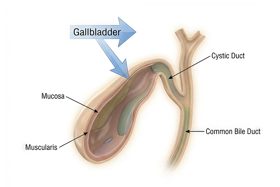
Functional gallbladder problems are another category ultrasound misses entirely. Understanding how the gallbladder functions as a contractile organ – filling with bile from the liver and squeezing it into the small intestine during digestion – makes it clear why sound-wave imaging cannot measure that process.
When the gallbladder fails to empty properly, the problem shows up in symptoms, not in structure. That requires a different kind of test entirely.
When a CT Scan for Gallbladder Disease Is the Right Next Step
When abdominal pain is severe, worsening, or accompanied by fever, jaundice, or signs of infection, physicians typically move to a CT scan for gallbladder disease evaluation without waiting for a second ultrasound. CT provides a complete cross-sectional view of the entire abdomen and pelvis – capturing not just the gallbladder but every adjacent structure that could be contributing to the problem.
A full abdominal CT scan adds several diagnostic capabilities that ultrasound cannot match. The comparison below outlines which modality fits which clinical scenario.
| Clinical Scenario | Best Imaging Choice | Why |
|---|---|---|
| Suspected uncomplicated gallstones | Ultrasound (first-line) | High sensitivity for stones; no radiation; fast and widely available |
| Mild acute cholecystitis | Ultrasound (first-line) | Detects wall thickening and pericholecystic fluid adequately in most patients |
| Suspected gangrenous or emphysematous cholecystitis | CT scan | CT detects gas in the gallbladder wall and other perforation-related signs ultrasound misses |
| Gallbladder perforation or suspected abscess | CT scan | CT maps the full extent of infection and guides surgical planning |
| Possible gallstone pancreatitis | CT scan or MRI | CT evaluates the pancreas; MRCP finds bile duct stones with superior detail |
| Pain with fever, jaundice, and rigors (Charcot's triad) | CT scan (urgent) | Must evaluate for cholangitis, biliary obstruction, or septic source |
| Inconclusive ultrasound with persistent symptoms | CT scan or HIDA scan | CT rules out other causes; HIDA scan evaluates gallbladder contractile function |
| Suspected bile duct stones without visible duct dilation | MRI with MRCP | MRCP has superior sensitivity for choledocholithiasis and avoids ionizing radiation |
Research published through the National Institutes of Health confirms that CT outperforms ultrasound for detecting gallbladder complications, with sensitivity for gangrenous cholecystitis reaching 90% or higher on CT compared to the 20-50% range reported for ultrasound in high-risk patients.
Decisions around whether a CT scan uses contrast material also matter in this context. For gallbladder complications, intravenous contrast helps highlight inflamed tissue, identify vascular compromise, and delineate abscess walls – features that non-contrast CT can miss in ambiguous presentations.
Other Imaging Options When a Gallbladder Ultrasound Is Not Enough
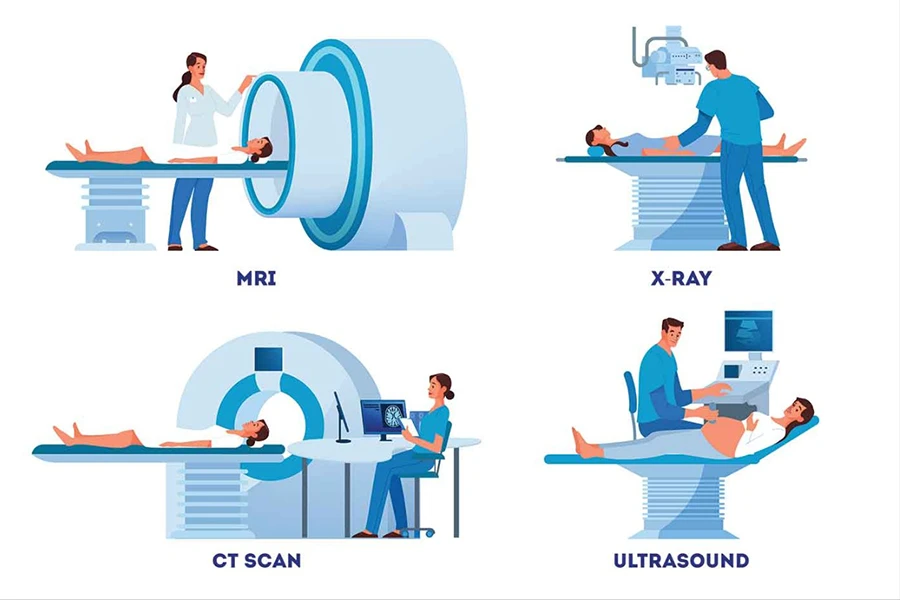
When both ultrasound and CT fall short of a clear diagnosis, two additional tools enter the picture. Each addresses a specific diagnostic gap that the first two modalities cannot fill.
The HIDA scan (hepatobiliary iminodiacetic acid scan) uses a small amount of radioactive tracer injected intravenously to measure how well the gallbladder fills with bile and empties during a simulated meal stimulus. It is the definitive test for biliary dyskinesia and acalculous cholecystitis – conditions where the gallbladder malfunctions without visible stones or obvious structural abnormalities on cross-sectional imaging. A gallbladder ejection fraction below 35% is generally considered abnormal and often guides the decision to proceed with cholecystectomy in patients whose symptoms align.
MRI with MRCP (magnetic resonance cholangiopancreatography) provides detailed images of the bile ducts, pancreatic duct, liver, and gallbladder without any ionizing radiation exposure. It excels at finding small bile duct stones that CT misses, differentiating gallbladder polyps from early tumors, and evaluating complex biliary anatomy before surgery. For patients who cannot receive CT contrast dye, or in whom bile duct stones are strongly suspected after a nondiagnostic ultrasound, MRCP is frequently the preferred escalation path.
Choosing between these tools comes down to the clinical question being asked. Structural pathology – perforation, abscess, pancreatitis – calls for CT. Functional pathology – poor gallbladder emptying, dyskinesia – calls for HIDA.
Complex biliary anatomy or small duct stones call for MRCP. Knowing which question you are actually trying to answer points to which scan to request.
Clinical Insight
Studies suggest up to 40% of patients with biliary dyskinesia have a completely normal gallbladder ultrasound on initial evaluation. Persistent right upper quadrant pain after a "normal" scan is not a closed case - it is a signal that functional imaging may be the missing piece.
What an Abdominal CT Scan Reveals Beyond the Gallbladder
One underappreciated advantage of CT imaging for gallbladder pain is the scope of what it evaluates simultaneously. A gallbladder ultrasound targets the gallbladder and bile ducts specifically. A CT scan of the abdomen captures the pancreas, liver, kidneys, bowel, adrenal glands, and abdominal vasculature in a single pass – often revealing pathology that had nothing to do with the original suspicion.
Abdominal pain is notoriously nonspecific. Right upper quadrant pain that presents like a gallbladder problem sometimes turns out to be a hepatic lesion, a duodenal ulcer, or an incidental pancreatic finding.
Understanding what a CT scan detects in a single abdominal study often surprises patients who expected to learn about only one organ. The scan does not limit itself to the chief complaint.
Pain in the right lower abdomen that migrates upward can point to appendicitis imaging territory. Left-sided discomfort that radiates toward the right flank can suggest diverticulitis imaging as the more likely diagnosis.
CT evaluates all of it in a single acquisition. And for patients who have a history of kidney stones, imaging for kidney stones and gallbladder disease can be addressed in the same CT study since the scan captures the entire urinary tract alongside the biliary system.
For patients who have been dealing with recurring abdominal symptoms without a confirmed cause, the panoramic view that abdominal CT provides often connects dots that individual organ-targeted studies have left scattered across multiple appointments and inconclusive reports.
When to Consider Proactive Abdominal Imaging
Not all gallbladder disease announces itself with an acute pain crisis. Many gallstones are silent – discovered incidentally on imaging ordered for an unrelated reason – and early gallbladder polyps rarely cause symptoms until they grow. The challenge is that the gallbladder sits alongside organs that carry serious long-term consequences if pathology goes undetected.
Craft Body Scan's full body scan includes abdominal imaging that evaluates major organs without a physician referral or insurance gatekeeping. For patients with a family history of gallbladder disease, a prior episode of gallstones, or chronic right-sided discomfort that has never been formally investigated, proactive abdominal screening provides the kind of visibility that symptom-triggered imaging – ordered only when pain becomes severe – cannot offer.
The virtual colonoscopy service at Craft Body Scan also evaluates the abdominal cavity as part of its standard protocol, capturing incidental abdominal findings beyond the colon during the same study. For patients concerned about multiple abdominal systems, combining scans during one appointment is an efficient approach that does not require multiple referrals or multiple sessions. Review scan pricing and packages to see what each study covers and how scans can be paired.
Frequently Asked Questions
Can a gallbladder ultrasound miss gallstones?
Yes, under certain conditions. Stones smaller than 2mm may fall below the resolution threshold of standard ultrasound transducers. Stones impacted in the cystic duct can also evade detection because they sit outside the primary field of view for the gallbladder body.
Additionally, a gallbladder that is not adequately distended – because the patient ate within 4-6 hours before the study – reduces stone visibility significantly. Fasting before the exam improves accuracy, which is why most facilities require it. If the study was technically limited due to body habitus or patient positioning, the radiologist's report typically notes it; that caveat is worth discussing with the ordering physician.
What does it mean when a gallbladder ultrasound is normal but pain continues?
A normal gallbladder ultrasound in the presence of continuing right upper quadrant pain typically signals that the problem is functional, structural but not yet visible, or located in an adjacent organ. It does not exclude biliary dyskinesia, acalculous cholecystitis, bile duct stones, or referred pain from the liver, pancreas, or bowel.
The next steps commonly include a HIDA scan to assess contractile function, an MRI with MRCP for bile duct evaluation, or a CT scan to broaden the diagnostic scope beyond the gallbladder itself. Accepting a normal ultrasound as a final answer when pain persists is one of the most common ways gallbladder disease gets delayed.
How does a CT scan differ from a gallbladder ultrasound?
A gallbladder ultrasound uses sound waves and evaluates the gallbladder and bile ducts specifically. A CT scan uses X-ray technology to image the entire abdomen and pelvis in cross-section, typically in a matter of seconds. CT provides superior detection of gallbladder complications including perforation, gangrenous change, abscess, and simultaneous pathology in adjacent organs.
The tradeoff is ionizing radiation exposure, which ultrasound avoids entirely. In emergency settings or when complications are suspected, CT is almost universally preferred because the diagnostic scope it provides cannot be matched by a targeted organ study.
When should I ask for a CT scan for gallbladder pain?
Consider asking about CT imaging when pain is severe, worsening over 24 to 48 hours, accompanied by fever above 101 degrees F, or associated with jaundice or vomiting that does not resolve with conservative management. These signs suggest a complication – gangrenous cholecystitis, perforation, or biliary abscess – that ultrasound is not sensitive enough to reliably exclude.
If your gallbladder ultrasound came back normal but symptoms have persisted beyond one week without improvement, a CT scan or HIDA scan is a reasonable next step to raise with your physician. You should not have to wait for pain to become unbearable before escalating imaging.
Is a HIDA scan better than a gallbladder ultrasound?
They are measuring fundamentally different things, so the comparison is not about which is better – it is about which question needs answering. A gallbladder ultrasound asks whether there are stones or visible structural problems. A HIDA scan asks whether the gallbladder is contracting and emptying properly.
For patients with persistent pain and a normal ultrasound, a HIDA scan is often the missing piece of the diagnostic picture. It is not a replacement for ultrasound; it is a functional complement to structural imaging that addresses what sound waves cannot see.
Can gallbladder disease be detected in a full body scan?
Yes. A full body CT scan evaluates the abdominal organs – including the gallbladder, liver, and bile ducts – as part of its standard imaging protocol. Gallstones, gallbladder wall abnormalities, biliary duct dilation, and adjacent organ pathology are all visible on abdominal CT imaging.
For patients who want full abdominal evaluation alongside cardiac, pulmonary, and other organ screening, Craft Body Scan offers direct-access scanning without requiring a physician referral. For more on what abdominal imaging reveals beyond targeted studies, the hernia imaging guide covers adjacent findings that frequently surface alongside gastrointestinal complaints.
Take the Next Step
A gallbladder ultrasound is a starting point – not a final answer. For patients who receive a normal or inconclusive result while symptoms persist, the path forward is to pursue imaging that matches the complexity of what the body is signaling.
CT scans, HIDA scans, and MRI each fill specific gaps that ultrasound leaves open. The right combination depends on symptom pattern, duration, and clinical severity – and no patient should have to settle for a diagnosis of "nothing found" when pain keeps returning.
Craft Body Scan offers direct-access abdominal imaging that patients can schedule without a physician referral. Board-certified radiologists review every scan, and results are delivered in language you can act on. If you have been dismissed after an inconclusive gallbladder ultrasound and want a clearer picture of what your abdominal organs are doing, take control of the process.
Ready for Real Answers?
Schedule your abdominal scan today. Radiologist-reviewed results. No referral needed.
Book Your Scan