
An ovarian cyst on ultrasound is typically the first image a clinician reviews when something surfaces during a routine pelvic exam or a scan ordered for pelvic pain. For most women, that image settles the matter quickly. But for some – when the cyst has an unusual shape, internal architecture, or features that don't fit a clean category – the question shifts from "is something there?" to "what kind of something is this?" That second question is where ultrasound and MRI diverge sharply.
Ultrasound is fast, widely available, radiation-free, and the standard first-line tool for evaluating ovarian cysts. It captures size, wall thickness, internal fluid, and structural features in real time. What it cannot reliably do is confirm tissue type, identify subtle hemorrhage with certainty, or distinguish particular complex cysts from early ovarian malignancy.
The findings on an ovarian cyst on ultrasound are always a starting point – not a final answer.
MRI fills some of those gaps – higher soft-tissue contrast, better characterization of cyst contents, and no blind spots created by bowel gas or body habitus. But MRI is not the final word either.
Neither modality replaces a tissue diagnosis. Both serve a specific role in a clinical pathway that most women with an ovarian cyst never need to travel very far down.
This guide walks through what each imaging tool actually reveals, where each one stops short, and what it means to find an ovarian cyst through proactive preventive scanning.
How Ultrasound Images an Ovarian Cyst
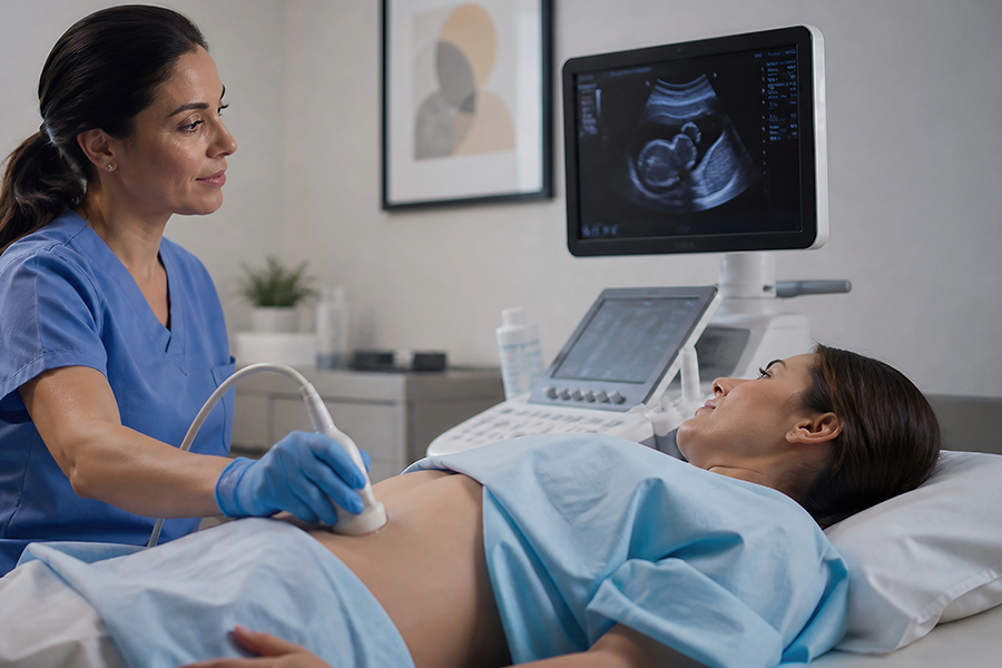
During a pelvic ultrasound – and more specifically, a transvaginal ultrasound, which places the probe near the cervix for sharper resolution – sound waves pass through tissue and reflect off structures with different densities. The cyst’s outer wall, its fluid contents, and any internal architecture all return distinct echoes that compose the image a radiologist reads.
Five features drive that reading.
- Wall thickness tells the radiologist whether the cyst boundary is smooth and thin or thickened and irregular.
- Interior appearance – described as anechoic when dark and fluid-only, or echogenic when internal reflections appear – signals the contents.
- Septations are internal dividing walls some cysts develop; they raise or lower concern depending on their number and thickness.
- Solid components such as nodules attached to the interior wall increase scrutiny.
- Doppler vascularity maps blood flow to indicate whether any solid area is receiving a vascular supply – a feature that carries clinical significance.
For most women of reproductive age, this single scan is enough. Simple, anechoic cysts under 3 centimeters are extremely common – many form and resolve naturally with the menstrual cycle – and rarely require anything further. For women pursuing proactive health screening, knowing that baseline makes the rest of this discussion more useful, not more alarming.
Simple vs Complex Ovarian Cyst on Ultrasound – What Each Type Means
The terms “simple” and “complex” do more clinical work than they might suggest. They are not judgments about severity. They are descriptions of what the image shows – and those descriptions drive the next step.
A simple ovarian cyst on ultrasound presents as a single, round or oval fluid-filled structure with a thin smooth wall and no internal echoes, septations, or solid components. These cysts carry a malignancy risk that research consistently places well below 1% in premenopausal women. Most resolve without intervention.
A complex ovarian cyst contains at least one additional feature – thickened or irregular walls, internal debris, multiple septations, or solid nodules.
The word “complex” does not mean malignant. The majority of complex cysts are benign. It means the image requires more context.
| Feature | Simple Ovarian Cyst | Complex Ovarian Cyst |
|---|---|---|
| Wall appearance | Thin, smooth | Thickened, irregular, or papillary projections |
| Interior | Anechoic (dark, fluid-only) | Internal echoes, debris, or solid components |
| Septations | None | Present - thin or thick |
| Malignancy risk | Very low (<1% in reproductive-age women) | Low to moderate depending on specific features |
| Typical next step | Follow-up ultrasound or no action | MRI, CA-125 blood test, or specialist referral |
| Common subtypes | Follicular cyst, corpus luteum cyst | Endometrioma, dermoid cyst, hemorrhagic cyst |
Certain subtypes carry a recognizable ultrasound signature. Endometriomas typically show a diffuse ground-glass echogenicity. Dermoid cysts contain fat and calcification that produce bright, echogenic foci with posterior acoustic shadowing.
Hemorrhagic cysts display a lace-like internal reticular pattern as the blood organizes. Each of these can be suggested by ultrasound – but none can be confirmed by imaging alone. Understanding the difference between a cyst and a tumor starts with the imaging characteristics and ends with pathology.
The Limits of Ultrasound in Ovarian Cyst Evaluation
Ultrasound was designed to detect structure. It does that exceptionally well. What it was not designed to do is determine tissue composition at the cellular level – and that limitation matters when the clinical question changes from “is there a cyst?” to “what is this cyst made of?”
Image quality itself introduces variability. Body habitus, bowel gas, and patient positioning can degrade resolution enough that an ovarian cyst on ultrasound may be noted as “not adequately evaluated” rather than classified. When a technically limited scan produces an inconclusive result, the radiologist’s report says so – and MRI becomes the next logical step.
An abnormal or indeterminate imaging result is not a diagnosis. It is a routing instruction.
Doppler vascularity adds information but not certainty. Blood flow to a solid component is a meaningful finding. It is not proof of malignancy.
Ultrasound stratifies probability. That stratification is valuable – but it is not the same as identification.
What MRI Adds to Ovarian Cyst Evaluation
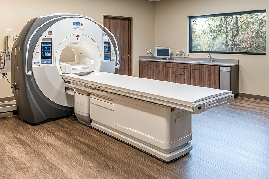
MRI generates images based on the hydrogen content of different tissues using magnetic fields and radiofrequency pulses. That mechanism produces soft-tissue contrast that ultrasound cannot approach – and that contrast is exactly what indeterminate ovarian cyst findings need.
A hemorrhagic cyst that appeared ambiguous on ultrasound produces a distinctive bright T1 signal on MRI that is hard to misread. An endometrioma shows characteristic “T2 shading” that reflects organized blood products layered over time. A dermoid cyst contains fat that MRI identifies definitively through fat-suppression sequences – a feature ultrasound can only suggest.
When a solid component carries Doppler blood flow, some pelvic MRI protocols also use gadolinium contrast to map vascular enhancement – one of the most common questions patients ask before an MRI is ordered. These characteristics narrow the differential diagnosis in ways that change clinical management.
MRI also covers the entire pelvis without the technical limitations of transvaginal ultrasound. Adjacent structures – uterus, fallopian tubes, pelvic lymph nodes, pelvic sidewall – are all visible, giving the radiologist a broader view of whether a cyst’s influence extends beyond the ovary itself. The role MRI plays in early detection of soft-tissue abnormalities goes well beyond ovarian cysts, but the ovaries are among the structures that benefit most from its soft-tissue contrast.
| Capability | Ultrasound | MRI |
|---|---|---|
| First-line use | Yes - standard initial tool | No - used when ultrasound is inconclusive |
| Radiation | None | None |
| Soft-tissue contrast | Moderate | High - definitive for tissue characterization |
| Identifies simple vs complex | Yes - primary classification tool | Yes - often more definitive |
| Detects hemorrhage | Limited - reticular pattern only | Strong - T1 bright signal |
| Confirms fat (dermoid) | Partial - echogenic foci suggest it | Definitive - fat-suppression sequences |
| Pelvic field of view | Limited by probe position and bowel gas | Full pelvis, no field-of-view limitations |
| Confirms malignancy | No - only biopsy/pathology does | No - only biopsy/pathology does |
The Malignancy Question – What Imaging Can and Cannot Confirm

This is the question most women are actually asking when they search for information about an ovarian cyst on ultrasound. Imaging identifies features that raise or lower the probability of malignancy.
It cannot, by itself, confirm or rule out cancer. Only a tissue biopsy examined by a pathologist does that.
What imaging does well is stratify risk. A simple, thin-walled, anechoic ovarian cyst on ultrasound carries such a low likelihood of malignancy that no further workup is typically needed. A complex cyst with Doppler-confirmed blood flow to a solid nodule warrants referral to a gynecologic oncologist.
MRI can push that stratification further for borderline cases – characterizing ambiguous findings more precisely – but the output is still a probability, not a verdict.
Key Context
According to the National Cancer Institute, the lifetime risk of ovarian cancer is approximately 1 in 78 women. Ovarian cysts - including complex ones - are far more common than ovarian cancer, and the imaging pathway exists precisely to identify the rare cases that need closer attention. The goal is not to alarm. It is to route the right cases to the right level of care.
The American College of Radiology's O-RADS (Ovarian-Adnexal Reporting and Data System) scoring system now provides a standardized framework for classifying ovarian cysts by malignancy risk on ultrasound. Scores range from 1 (no cyst, physiologic finding) to 5 (high risk features). MRI has its own O-RADS scoring extension.
Together, these systems give radiologists a shared vocabulary that reduces variability in how complex findings are reported and followed. Research published through the National Institutes of Health supports O-RADS as a tool that measurably reduces unnecessary referrals for low-risk cysts. Connecting those findings to the right cancer screening pathway is where clinical management begins.
Preventive MRI Scanning and Women’s Pelvic Health

Most ovarian cysts are found because a woman has symptoms – pelvic pain, bloating, or an irregular exam – that prompt targeted imaging. A growing number of women find them another way. A whole-body MRI scan covers abdominal and pelvic structures as part of its field of view, and ovarian findings – simple cysts, complex cysts, and other adnexal structures – can appear as incidental findings even when pelvic imaging was not the primary purpose.
This is not a replacement for targeted transvaginal ultrasound when symptoms or clinical concerns direct one. It is a different entry point – a broader sweep that places pelvic anatomy into the context of the full abdomen and chest. Women who pursue full body preventive screening sometimes get their first look at structures they had never previously imaged.
Any finding on a whole-body MRI reviewed at Craft Body Scan is read by a board-certified radiologist, who notes whether follow-up or further evaluation is recommended.
The full range of preventive imaging services available at Craft Body Scan – including MRI, CT, and ultrasound-based options – covers multiple organ systems in a single visit. For women with a family history of ovarian cancer or gynecologic conditions, that broader view is worth factoring into a preventive health plan – alongside other women’s cancer screening priorities a preventive imaging plan can address.

Frequently Asked Questions
What does an ovarian cyst look like on an ultrasound?
A simple ovarian cyst on ultrasound appears as a round or oval dark structure with a thin, smooth wall and a clear interior. That dark interior – called anechoic – reflects the fluid inside, which does not return sound waves the way solid tissue does. A complex cyst looks more varied: internal echoes, irregular walls, visible septations, or attached solid components create a more detailed image that requires closer interpretation.
Can ultrasound tell if an ovarian cyst is cancerous?
Ultrasound identifies features that raise or lower the probability of malignancy – wall irregularity, solid components, Doppler blood flow. It cannot confirm cancer. That determination requires pathology from a tissue sample.
Ultrasound's role is to classify the cyst's imaging characteristics and route borderline or high-concern findings to the appropriate next step, whether that means MRI, a CA-125 blood test, or specialist referral.
When is MRI ordered for an ovarian cyst instead of ultrasound?
MRI is typically ordered when an ovarian cyst on ultrasound produces indeterminate findings – a complex structure that the ultrasound could not clearly classify as benign or concerning. It is also used when body habitus or bowel gas limited ultrasound quality, when a specific cyst subtype (such as endometrioma or dermoid) needs confirmation, or when a clinician needs detail about adjacent pelvic structures that ultrasound cannot adequately cover.
What is the difference between a simple and complex ovarian cyst on imaging?
A simple cyst has one chamber, a thin smooth wall, and no internal architecture. A complex cyst has at least one additional feature – thickened walls, internal echoes, septations, or solid nodules. Most complex cysts are benign.
The classification drives the clinical pathway, not the diagnosis. According to RadiologyInfo.org, the large majority of ovarian cysts found during pelvic ultrasound are non-cancerous and require only monitoring.
Can a whole-body MRI detect ovarian cysts?
Yes. Ovarian cysts are visible on the pelvic sequences included in a whole-body MRI. Women who undergo a whole-body preventive scan may have ovarian findings identified as incidental discoveries – findings they did not specifically scan for but that appear within the MRI's field of view.
Every scan at Craft Body Scan is reviewed by a board-certified radiologist, who notes any finding that warrants follow-up attention.
Take the Next Step
Most ovarian cysts resolve without intervention. The imaging pathway exists for the ones that need a second look – and knowing what ultrasound reveals, what MRI adds, and where both tools stop short gives women the foundation to move through that pathway with clarity rather than uncertainty.
Early detection works best when you already have a baseline. Waiting for symptoms is one approach. Building a picture of your health before symptoms appear is another.
Craft Body Scan's board-certified radiologists review every scan result. Whether you are booking your first preventive imaging appointment or following up on a prior finding, take control of your health with early detection.
Ready to Take Control of Your Health?
Craft Body Scan offers whole-body MRI, full body scans, and preventive imaging services reviewed by board-certified radiologists - no referral required.
Schedule Your Scan