You got your PSA results back. The number is elevated. Your doctor mentioned imaging – or possibly a biopsy – and now you are trying to understand what that means and what comes next.
This is where most patients hit a wall. PSA is just a number. The path from that number to a clear action plan is rarely explained well. High PSA next steps depend on more than the absolute value – they depend on your age, how fast your PSA has risen, the size of your prostate, and whether imaging can clarify what a biopsy alone cannot. If you are already tracking your cancer screening options, imaging after an elevated PSA is one of the most important steps you can take.
Most clinical guidelines now recommend a multiparametric MRI (mpMRI) of the prostate when PSA is elevated – and often before a biopsy is performed. But the threshold that triggers imaging is not a single cutoff. It shifts depending on whether your PSA sits in the gray zone (4-10 ng/mL), is clearly elevated (above 10 ng/mL), or is rising faster than expected over time. This article walks through the PSA levels that typically trigger imaging, the type of scan used, how results are scored, and what the pathway forward looks like.
How PSA Levels Guide the Decision to Image

Prostate-specific antigen is a protein produced by the prostate gland. Elevated PSA signals that something has changed – but not necessarily cancer. Benign prostatic hyperplasia (BPH), prostatitis, and even a recent digital rectal exam can push the number higher. That is exactly why imaging exists as a middle step between an abnormal lab result and a tissue biopsy.
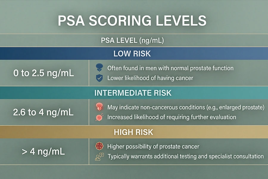
To understand what constitutes a dangerous PSA level, context matters. Age-adjusted reference ranges are the starting point. Once a level is flagged as elevated for your age group, the next question is what action that warrants. Normal PSA levels by age vary significantly – a result of 4.5 ng/mL reads differently in a 45-year-old than in a 70-year-old. What follows is how clinicians typically decide whether imaging is the right next step.
| PSA Range (ng/mL) | Typical Clinical Response | When Imaging Is Considered |
|---|---|---|
| Below 4 | Generally within normal range (age-adjusted) | Not typically indicated unless PSA velocity is high |
| 4 - 10 (Gray Zone) | Elevated - further evaluation recommended | mpMRI often recommended before biopsy decision |
| 10 - 20 | Clearly elevated - significant cancer risk signal | mpMRI strongly recommended prior to biopsy |
| Above 20 | Very high - aggressive workup indicated | Imaging ordered alongside biopsy planning |
| Any level - rising velocity | PSA rise of more than 0.75 ng/mL per year | Imaging recommended regardless of absolute value |
PSA density adds another layer to this picture. It is calculated by dividing PSA by prostate volume – and a density above 0.15 is treated as a higher-risk signal even when the absolute number sits in the gray zone. A large prostate producing more PSA from benign tissue tells a different story than a small prostate with the same number. Understanding how the prostate functions helps put both of these metrics in context.
What Type of Imaging Is Used After High PSA
Not all imaging serves the same purpose when it comes to prostate evaluation. The tool that has become the clinical standard is multiparametric MRI – often written as mpMRI or mp-MRI. It combines three MRI sequences to give radiologists a detailed look at prostate tissue from multiple angles simultaneously.
According to the American College of Radiology, mpMRI is now recommended by major urology guidelines before performing a prostate biopsy in patients with elevated PSA – particularly those in the gray zone. The rationale is direct. Targeted imaging helps identify suspicious regions so that any biopsy can be guided toward the area of concern, rather than sampling tissue at random.
| Imaging Type | What It Shows | Role in PSA Workup |
|---|---|---|
| Multiparametric MRI (mpMRI) | Prostate zones, suspicious lesions, tissue characteristics | First-line imaging after elevated PSA - guides biopsy targeting |
| Transrectal Ultrasound (TRUS) | Prostate size, basic structural changes | Often used to guide biopsy needle placement |
| Whole Body MRI | Soft tissue organs including prostate, lymph nodes, and other organ systems | Useful for broad cancer screening and staging review |
| Bone Scan (Nuclear Medicine) | Bone metastases | Used when PSA is very high and metastatic disease is suspected |
| PSMA PET Scan | Cancer cells expressing PSMA protein | Recurrence detection after treatment - not a first-line PSA workup tool |
The free PSA ratio is another factor that shapes the imaging decision. When the percentage of free PSA in the blood is low (typically below 10-25%), the likelihood of cancer being the driver behind an elevated number increases. Physicians weigh this ratio alongside PSA velocity and prostate volume before finalizing whether imaging is the appropriate next move.
How mpMRI Results Are Scored

When a prostate MRI is completed, the radiologist assigns a PI-RADS score to any finding identified. PI-RADS stands for Prostate Imaging – Reporting and Data System. The scale runs from 1 to 5 and communicates the likelihood that a lesion represents clinically significant cancer.
PI-RADS Score Reference
- PI-RADS 1 - Very low: No significant lesion identified. Standard follow-up with your physician.
- PI-RADS 2 - Low: No significant finding. Continued PSA monitoring is recommended.
- PI-RADS 3 - Intermediate: Indeterminate. Additional workup or targeted biopsy may follow depending on PSA density and clinical profile.
- PI-RADS 4 - High: Findings suspicious for cancer. Targeted biopsy is typically recommended.
- PI-RADS 5 - Very high: Highly suspicious for clinically significant cancer. Biopsy is recommended without delay.
Research published through the National Library of Medicine supports mpMRI-guided biopsy as significantly more accurate than random biopsy alone – particularly for detecting clinically significant prostate cancer while reducing detection of low-grade disease that may not require treatment. Finding what matters, and not chasing what does not, is the core advantage of imaging-first protocols.
A PI-RADS score below 3 does not mean the situation is ignored. Your physician will factor in your PSA trend, your free PSA ratio, your prostate volume, and your family history. The score gives context – it does not replace the clinical conversation.
What Happens After Prostate MRI – The Pathway Forward
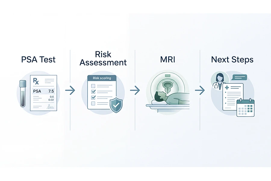
Once imaging is complete and scored, your urologist has a much clearer picture to work from. The pathway that follows depends on where the PI-RADS score lands and what your broader risk profile looks like.
- PI-RADS 1 or 2 results typically move patients to active surveillance. PSA testing continues on a schedule – often every 6-12 months. No biopsy is indicated unless PSA rises significantly or velocity accelerates.
- PI-RADS 3 results represent a judgment call. Your physician may recommend a targeted biopsy, a repeat MRI at 6-12 months, or further risk stratification using free PSA ratio or other biomarker tests.
- PI-RADS 4 or 5 results typically lead to a targeted biopsy using MRI-ultrasound fusion guidance. This approach combines the MRI’s lesion map with real-time ultrasound to sample the specific area of concern – not random tissue.
- Very high PSA or suspicious lymph nodes on MRI may prompt staging scans – including bone scans or PSMA PET imaging where available – to assess whether cancer has spread beyond the prostate.
This process is stepwise, not rushed. The goal of imaging is to avoid unnecessary biopsies in men with low-risk findings while ensuring that high-risk findings get timely tissue confirmation. Knowing your high PSA next steps before the appointment means you walk in prepared to ask the right questions. According to the National Cancer Institute, early detection of prostate cancer – when disease is still localized – is associated with significantly better treatment outcomes.
Other Factors That Shape the Imaging Decision
A single elevated PSA number rarely tells the complete story. Physicians look at a combination of variables when deciding whether imaging is the right next step or whether watchful waiting makes more sense.
- PSA velocity matters as much as the absolute number. A rise of more than 0.75 ng/mL per year is considered clinically meaningful – even if the absolute level stays within a technically normal range for that age group.
- Free PSA ratio helps distinguish BPH from cancer. When the percentage of free PSA is lower (below 10-25%), the likelihood of cancer as the driver increases.
- Prostate size affects PSA density. A large prostate producing more PSA from benign tissue tells a different story than a small prostate with the same PSA level.
- Age and overall health factor into whether aggressive workup is appropriate. Clinical priorities differ for a 75-year-old with significant comorbidities versus a 55-year-old who is otherwise healthy.
- Family history of prostate cancer – particularly a first-degree relative – increases baseline risk and often lowers the threshold at which imaging is ordered.
When these factors combine unfavorably – rising PSA velocity, low free PSA ratio, family history, a smaller prostate volume that makes density high – imaging becomes the obvious next step even before the absolute PSA number crosses a hard threshold. That is the clinical reality behind the phrase “high PSA next steps.” There is no single number. There is context.
Where Craft Body Scan Fits in Your Prostate Health Journey
For men weighing their high PSA next steps and wondering where to begin, Craft Body Scan’s AI-powered whole body MRI scan gives board-certified radiologists a detailed view of soft tissue organs throughout the body – including the prostate, surrounding lymph nodes, and other areas that can signal early disease. It is not a replacement for a dedicated prostate mpMRI ordered through urology, but it is a meaningful tool for men who want a broader view of what is happening inside before symptoms appear.
If you are monitoring PSA, have a family history of prostate cancer, or simply want to know what a full body scan can detect in terms of prostate and organ health, Craft Body Scan provides that picture without requiring a physician referral. Board-certified radiologists review every scan and findings become part of the conversation you have with your doctor about next steps.
For men at elevated risk who want imaging that goes beyond a single organ, Craft Body Scan’s full body scan options are designed around early detection – the point in time when action is most effective. If you have been exploring early detection with MRI as part of a proactive health approach, this is where that conversation starts.

Frequently Asked Questions
What PSA level triggers an MRI?
Most urologists consider mpMRI for PSA in the gray zone of 4-10 ng/mL when a biopsy is being evaluated. For PSA above 10 ng/mL, imaging is strongly recommended before biopsy. High PSA next steps almost always include imaging at this threshold. PSA velocity – how fast the number is rising year over year – can also trigger imaging even when the absolute level appears borderline for a given age.
Can I skip the biopsy if my MRI comes back low risk?
If your mpMRI scores PI-RADS 1 or 2, your physician may recommend active surveillance instead of an immediate biopsy. This means monitoring PSA over time rather than pursuing tissue sampling right away. The decision depends on your age, PSA trend, free PSA ratio, and overall health – and is always made in partnership with your urologist, not unilaterally.
Is a whole body MRI the same as a prostate MRI?
No. A whole body MRI scans multiple organ systems simultaneously, including soft tissue around the prostate and pelvic lymph nodes. A dedicated prostate mpMRI focuses specifically on the prostate using sequences optimized for tissue characterization and PI-RADS scoring. Both can reveal abnormalities. A whole body MRI is well suited for broad early detection screening, while a prostate mpMRI is used for diagnostic workup when PSA is elevated and biopsy guidance is needed.
Does an elevated PSA always mean cancer?
No. Elevated PSA is a signal, not a diagnosis. Benign prostatic hyperplasia (BPH), prostatitis, and vigorous physical activity can all raise PSA. Imaging and, if indicated, targeted biopsy are the tools that determine whether an elevated number reflects cancer or a non-cancerous condition. This is exactly why the imaging step exists before tissue sampling.
How long does it take to get prostate MRI results?
A dedicated prostate MRI typically takes 45-90 minutes to perform. Results are reviewed by a radiologist and usually returned to your ordering physician within 1-3 business days. Your urologist will then schedule a follow-up to walk through findings and outline next steps based on the PI-RADS score and your broader clinical picture.
How often should men get PSA tested?
Many physicians recommend annual PSA testing starting at age 50, or earlier – around age 40-45 – for men with a first-degree relative diagnosed with prostate cancer. The normal PSA range by age helps put each result in context. A number that warrants imaging at 45 may be entirely typical at 70, which is why age-adjusted reference ranges matter in every PSA conversation.
High PSA Next Steps – Take Control of Your Health
Elevated PSA results are not a verdict. They are a starting point. The pathway from an abnormal number to an accurate picture of your prostate health runs through imaging – specifically through an mpMRI that gives board-certified radiologists the detail needed to make informed recommendations. High PSA next steps work best when they begin early. The earlier imaging captures what is happening, the more options remain open.
Craft Body Scan exists for this moment – when you know something needs attention but you are not yet certain what that looks like. Whole body MRI, cancer screening packages, and a team of board-certified radiologists reviewing every scan. Take control of your health before symptoms arrive.
Ready to See What Your Scans Reveal?
Craft Body Scan offers whole body MRI and cancer screening at locations across Oklahoma, Texas, Florida, North Carolina, Tennessee, and Ohio - no referral required.
Schedule Your Scan