Medical note. This article is educational only. New or worsening belly pain can turn urgent, especially if you also have fever, vomiting, faintness, or a rigid abdomen. Call 911 or go to the ER instead of waiting for a scan appointment.
A bad diverticulitis flare and appendicitis can feel uncomfortably close at first. You may have belly pain, fever, nausea, and no useful clue from symptoms alone. The side of the pain helps, but diverticulitis vs appendicitis imaging usually makes the call. CT lets the radiologist check the appendix, colon, nearby fat, fluid pockets, perforation, and look-alike problems in one exam.
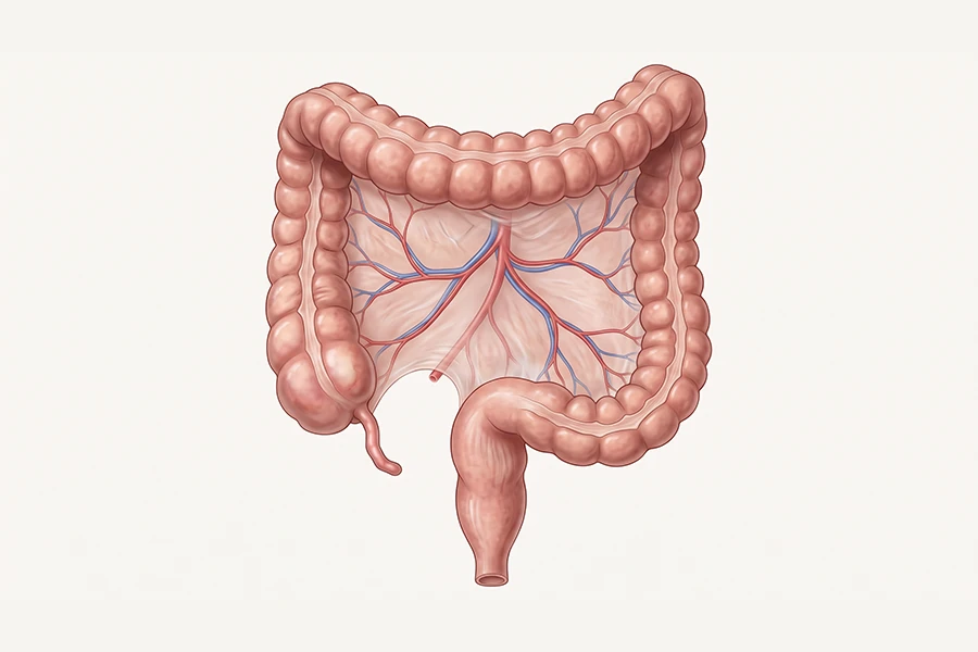
The anatomy is the real split. Appendicitis starts in the appendix, the small pouch attached to the large intestine on the lower right side. Diverticulitis starts in small pouches in the colon wall. In the U.S., those pouches usually sit in the sigmoid colon on the lower left side.
RadiologyInfo.org explains that abdomen and pelvis CT is commonly used for acute abdominal pain because it shows organs, bowel, blood vessels, bones, and soft tissue. When the symptom pattern points in two directions, our radiologists need that view.
If the pain is severe, started suddenly, or keeps getting worse, go to an emergency room. Read about the scan after you are safe.
The short answer
- Location gives the first clue because appendicitis usually hurts on the lower right. Diverticulitis more often hurts on the lower left, though either pattern can fool you.
- Age changes the odds because appendicitis is more common in younger adults. Diverticulitis rises after age 50, a core screening age for Craft Body Scan patients.
- CT is the main imaging test in adults when symptoms are serious or unclear. Research on appendicitis puts CT around 95% sensitivity and 94% specificity. Diverticulitis CT performance also lands in the mid-to-high 90s in published sources.
- Timing matters because either condition can become an emergency. After imaging confirms the problem, your care team may move quickly. Treatment can mean antibiotics, drainage, surgery, or a mix of those steps.
Diverticulitis vs appendicitis at a glance
Doctors start with this kind of comparison at the bedside. Imaging becomes necessary when the pattern is close enough to make treatment risky without a better look.
| Feature | Appendicitis | Diverticulitis |
|---|---|---|
| Typical age | Any age, with a peak from the teen years through the 30s | More common after age 50 and less common under 40 |
| Pain location | Lower right abdomen, also called the right lower quadrant | Lower left abdomen, though right-sided disease can happen |
| Onset speed | Often worsens within 24 hours | Often builds across 1 to 3 days |
| Fever and nausea | Common, with appetite loss often appearing early | Fever is common. Nausea can occur, but bowel changes may stand out more |
| Inflamed area | The appendix | Colon-wall pouches, usually in the sigmoid colon |
| Urgency | Rupture risk rises without care. Surgery is common | Antibiotics may work. Abscess or perforation may need drainage or surgery |
| Recurrence | Does not recur after appendectomy | Can recur, especially in people with known diverticular disease |
How the symptoms differ
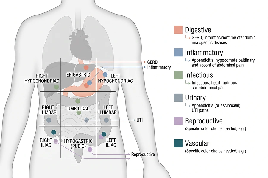
Pain location helps, but it cannot diagnose you by itself. Classic appendicitis often starts near the navel and then moves to the lower right abdomen. The tender exam spot is McBurney’s point, between the navel and the right hip bone.
Diverticulitis usually hurts on the lower left because the sigmoid colon sits there. That is the common Western pattern. Right-sided diverticulitis still happens, and older adults with appendicitis may not follow the textbook pain story. The bedside exam can only take the doctor so far.
Tenderness, guarding, nausea, fever, and a high white blood cell count can appear with either condition. Rebound tenderness can show up too. A doctor may suspect one diagnosis from the pattern, then use CT to check the anatomy.
Speed can help too. Appendicitis often moves fast. A normal morning can turn into severe pain by night. Diverticulitis may build more slowly, with constipation, diarrhea, or a few days of lower abdominal discomfort first.
Why imaging changes the diagnosis
The physical exam can miss more than patients expect. Appendicitis may look like gastroenteritis, kidney stones, gynecologic problems, or diverticulitis. Diverticulitis may mimic bowel symptoms, ovarian disease, urinary problems, or colorectal cancer. CT lets the radiologist read the anatomy instead of relying on pain location alone.
In adults with suspected appendicitis, a Cochrane review found CT sensitivity of 95% and specificity of 94%. The same review found IV contrast CT sensitivity of 96% and specificity of 93%. That is strong enough to make CT the main adult test when symptoms are serious or unclear.
CT sensitivity for appendicitis in adults (Cochrane review)
CT specificity for appendicitis in adults
Reported CT accuracy range for acute diverticulitis
Diverticulitis CT performance is strong too. Clinical references report CT sensitivity, specificity, and negative predictive value above 97% in some settings. Other radiology guidance puts sensitivity near 94% and specificity near 99%.
The number changes by protocol, patient factors, and study design. The practical point holds. A good CT study is highly reliable when the images and symptoms line up.
Ultrasound still has a role, especially for children and pregnant patients. The tradeoff is visibility. Bowel gas, body size, and appendix position can block the view. If ultrasound misses the appendix, doctors may still need CT or MRI depending on the patient.
Contrast choice depends on the question. IV contrast can help show bowel wall inflammation, abscesses, blood vessels, and abnormal contrast uptake. Some centers add oral contrast too. Kidney function, allergy history, pregnancy status, and scan urgency all affect the protocol. Our guide on CT scan with contrast vs no contrast explains how doctors choose.
What a CT scan shows for appendicitis
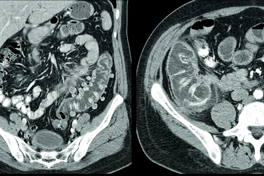
On CT, appendicitis is not diagnosed from size alone. A diameter over 7 millimeters, or over 6 millimeters in some protocols, matters more when it appears with surrounding inflammation. Radiologists look at the appendix, nearby fat, fluid, and signs of complication together.
- Appendix wall thickening appears when the wall becomes thicker and may light up after IV contrast, which points to active inflammation.
- Periappendiceal fat stranding means fat near the appendix turns hazy or streaky on CT when inflammation spreads outside the appendix wall.
- Appendicolith describes a small calcified blockage inside the appendix that can appear as a bright spot. Its presence may raise concern for a more complicated case.
- Free fluid or free air can change the urgency. Fluid near the appendix can occur with inflammation, while free air raises concern for perforation.
- Abscess formation can change timing and treatment planning because it shows a pocket of infected fluid near the appendix.
For many adults, CT with IV contrast gives surgeons and emergency physicians the clearest pre-treatment map. For more detail on this condition, our guide to appendicitis imaging explains the findings in a dedicated article.
What a CT scan shows for diverticulitis
Diverticulitis has a different CT pattern because the problem sits in the colon wall. In the United States, the inflamed segment is most often in the sigmoid colon, which runs through the lower left pelvis.
- Diverticula are small pouches along the colon wall. Those pouches show diverticulosis. Diverticulitis means the area around them is inflamed.
- Colon wall thickening appears in the affected segment where inflammation is active.
- Pericolic fat stranding shows hazy or streaky fat around the colon, similar to the pattern seen around an inflamed appendix.
- Abscess means infected fluid has collected near the colon. Treatment depends on size and severity and may include antibiotics, drainage, or surgery.
- Perforation signs include free air or contrast outside the colon, which can signal a leak through the bowel wall.
- Fistula formation can appear in severe or repeated cases when CT shows an abnormal connection from the colon to the bladder or another nearby organ.
A CT scan can detect diverticulitis with high accuracy because it shows the inflamed bowel segment and the tissue around it. Our full guide to diverticulitis imaging breaks down that workup in more detail.
When abdominal pain needs the ER
ER warning signs
- →Sudden severe abdominal pain that starts in minutes and does not improve.
- →A rigid or board-like abdomen when touched, which can point to peritonitis.
- →Fever above 101°F with meaningful abdominal pain.
- →Vomiting you can’t control along with lower abdominal pain.
- →Pain that keeps worsening across several hours.
- →Possible sepsis signs such as rapid heart rate, chills, confusion, or feeling faint.
Some abdominal pain is clearly acute. The warning signs above need an ER evaluation rather than a later appointment. Milder symptoms can be harder to judge, especially if you have a known history of diverticular disease or lower abdominal discomfort that comes and goes.
Urgent care centers with CT capability may handle mild to moderate cases. A primary care doctor can order imaging for non-emergency symptoms. For known diverticular disease, a gastroenterologist may guide care. If symptoms worsen, emergency evaluation is safer.
CT can also reveal causes that are not appendicitis or diverticulitis. Kidney stones, ovarian cysts, mesenteric lymphadenitis, bowel obstruction, and some cancers can cause similar symptoms. A useful CT read does more than name one diagnosis. It checks the nearby possibilities too. Our overview of 5 health problems a CT scan can find shows how much abdominal imaging can cover.
How preventive imaging fits in

Preventive abdominal imaging at Craft Body Scan is not emergency imaging. Severe pain today needs a hospital. If you feel well and want a clearer view of abdominal structures, a preventive scan can show anatomy that may deserve follow-up with your own doctor.
A board-certified radiologist reviews your scan and flags findings that warrant medical follow-up. Our full body scan includes abdominal imaging as part of the review. Patients focused on colon health can also review our colon imaging service. If cancer screening is part of your plan, our cancer screening options cover several organ systems in one visit.
The limitation matters. A preventive scan done when you feel fine is not meant to diagnose an active diverticulitis flare. Emergency CT protocols and preventive imaging protocols answer different questions. Preventive imaging is for structure, anatomy, and findings that may deserve planned follow-up. Our pricing page explains what is included. You can also read our guide to gallstone imaging if you are comparing abdominal concerns.
FAQ
How do you know if it’s diverticulitis or appendicitis?
The main clues are pain location, age, onset speed, appetite loss, fever, nausea, and bowel changes. Appendicitis more often causes lower right abdominal pain and can worsen quickly. Diverticulitis more often causes lower left abdominal pain and may build over days. The overlap is big enough that CT of the abdomen and pelvis is often needed when symptoms are moderate, severe, or unclear.
Can appendicitis be misdiagnosed as diverticulitis?
Yes, right-sided diverticulitis can feel similar to appendicitis, and older adults with appendicitis may not follow the classic pain pattern. CT is the most reliable way to separate the two in adults because it shows the appendix, colon, and surrounding tissue directly.
Can a CT scan detect diverticulitis?
Yes, CT is the main imaging test for suspected diverticulitis in many adult patients. It can show diverticula, colon wall thickening, pericolic fat stranding, abscess, perforation, and other causes of left lower abdominal pain. Reported accuracy is high, but the exact sensitivity varies by study and protocol.
What is the gold standard imaging for appendicitis?
For most nonpregnant adults, CT of the abdomen and pelvis is the main imaging test for suspected appendicitis. IV contrast is often preferred when kidney function and allergy history allow it. Ultrasound is often used first in children and pregnant patients. MRI may follow when ultrasound is unclear and radiation avoidance matters.
Will an ultrasound show appendicitis?
Sometimes, ultrasound can confirm appendicitis when the appendix is visible and inflamed. The challenge is that the appendix may be hidden by bowel gas, body habitus, or position. A non-visualized appendix on ultrasound does not always rule out appendicitis, so adult ER protocols often move to CT when symptoms still point that way.
Do I need contrast for an abdominal CT?
It depends on the clinical question and your health history. IV contrast can help show inflammation, abscess, blood vessels, and masses. Some protocols use oral contrast too. People with kidney disease or contrast allergy may need a different plan. In a preventive setting, the protocol is matched to the screening goal. Our guide to CT scan with contrast vs no contrast explains the decision in more detail.
Take the next step
Abdominal concerns that aren’t an emergency? Plan the next step.
If you have recurring abdominal discomfort, a history of diverticular disease, or questions about what is happening inside, a preventive abdominal scan at Craft Body Scan gives you a physician-readable report without a referral or ER visit. A board-certified radiologist reviews every scan and writes a report you can bring to your own doctor.