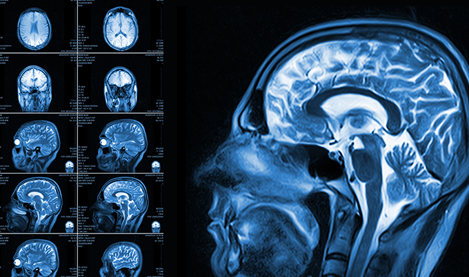
When a doctor orders a brain scan, the question that almost always follows is: “Why this one and not the other?” Understanding the difference between a brain MRI vs CT scan isn’t just medically interesting – it’s the kind of knowledge that helps you walk into a radiology appointment feeling prepared rather than anxious. These two technologies produce entirely different pictures of what’s happening inside your skull, and physicians choose between them for very specific reasons.
Both scans look at the brain. That’s where the similarity largely ends.
A CT scan uses X-ray beams rotating around the head to create cross-sectional images in minutes. It’s fast, widely available, and extraordinarily good at detecting certain emergencies – particularly bleeding, fractures, and acute stroke. An MRI uses powerful magnetic fields and radio waves to generate images with far greater soft tissue detail. It takes longer, costs more, and gives radiologists structural clarity that CT simply cannot match.
Which scan gets ordered shapes what gets found – and what gets missed.
Brain CT Scan: What It’s Designed to Find
Speed is the defining feature of a brain CT scan. In an emergency room, where every minute affects outcomes after a stroke or head injury, CT is usually the first imaging tool reached for. That urgency-driven design tells you a great deal about what CT does best.
A brain CT scan is particularly suited to detecting:
- Acute hemorrhage (bleeding in or around the brain) – Fresh blood appears bright white on CT, making it one of the most reliable ways to identify a hemorrhagic stroke or trauma-related bleeding.
- Skull fractures – CT captures bone with exceptional clarity, making it the standard tool for evaluating head injuries.
- Large tumors or masses – While MRI provides more detail on brain tumors, CT can identify significant masses, especially when contrast dye is used.
- Hydrocephalus (fluid buildup) – Enlarged ventricles and abnormal fluid accumulation show clearly on CT.
- Calcifications – Calcium deposits in brain tissue, which can signal certain conditions, are actually more visible on CT than MRI.
- Acute ischemic stroke – CT is typically the first scan run when stroke is suspected, to rule out bleeding before treatment begins.
CT is not the most sensitive tool for detecting early or subtle brain abnormalities. It’s the right tool for the right moment – emergencies, trauma, fast triage. For deeper investigation of soft tissue changes, the picture shifts significantly toward MRI.
Brain MRI vs CT Scan: What MRI Captures That CT Cannot
MRI is, by almost every measure, the more detailed imaging technology for brain soft tissue. The tradeoff is time – a standard brain MRI runs 30 to 60 minutes, compared to CT in under 10. But for conditions where that detail makes the difference between a finding and a miss, that time is worth it.
Brain MRI is the gold standard for evaluating:
- Multiple sclerosis (MS) and white matter disease – The demyelinating lesions characteristic of MS are often invisible on CT. MRI, particularly with FLAIR sequences, reveals them clearly.
- Brain tumors – MRI characterizes tumors by size, location, borders, and involvement of surrounding structures far better than CT. It’s the standard for surgical planning.
- Early or small strokes – Diffusion-weighted MRI sequences can detect ischemic stroke changes within minutes of onset, often catching damage CT would miss for the first 24-48 hours.
- Epilepsy and seizure evaluation – Subtle cortical abnormalities, hippocampal changes, and focal lesions associated with seizure activity are MRI territory.
- Infections and inflammation – Encephalitis, meningitis complications, and abscesses in brain tissue are evaluated with MRI, often with contrast.
- Vascular malformations – Cavernous malformations, arteriovenous malformations, and aneurysms (particularly small ones) are better characterized with MRI or MR angiography.
- Pituitary and posterior fossa abnormalities – The brainstem, cerebellum, and pituitary gland sit in areas where CT is limited by bone artifact. MRI sees these regions with clarity.
There’s a reason neurologists lean on MRI when investigating persistent headaches, cognitive changes, vision problems, or unexplained neurological symptoms. The resolution and tissue contrast MRI provides is in a different category for those applications.
When to Get a Brain CT Scan vs an MRI
The decision between a brain MRI vs CT scan usually isn’t arbitrary. There’s a clinical logic behind which one gets ordered, and understanding that logic helps make sense of what your doctor is – and isn’t – concerned about.
A brain CT scan is typically chosen when:
- The situation is urgent. Any presentation involving sudden severe headache, altered consciousness, suspected stroke, or head trauma gets a CT first. Speed outweighs detail in these moments.
- Bleeding needs to be ruled out fast. Before clot-dissolving medication like tPA can be given for stroke, doctors must confirm there’s no hemorrhage. CT provides that answer in minutes.
- The patient can’t tolerate an MRI. Metal implants, pacemakers, certain surgical clips, severe claustrophobia, or inability to lie still can all make CT the practical choice.
- Bone detail is the priority. Evaluating skull fractures or bony involvement of tumors is a CT strength.
Brain MRI is typically chosen when:
- A neurological condition needs thorough evaluation. New or progressive symptoms – cognitive decline, new seizures, unexplained weakness or numbness – warrant MRI’s detailed soft tissue imaging.
- A brain tumor has been identified or is suspected. MRI with and without contrast is the standard for characterizing, staging, and monitoring brain tumors.
- The CT came back normal but symptoms persist. A normal CT does not mean nothing is happening in the brain. Small strokes, early MS lesions, low-grade tumors – these can all show nothing on CT while appearing clearly on MRI.
- Monitoring a known condition. Patients with MS, brain tumors under treatment, or vascular malformations are typically followed with MRI over time.
Important: A normal CT result does not equal a normal brain MRI result. CT misses many conditions that MRI detects – including early MS, small strokes, and low-grade tumors. If symptoms persist after a normal CT, your physician may order an MRI as a next step. Learn more in our guide on what a normal brain MRI actually means (https://craftbodyscan.com/blog/normal-brain-mri/).
Radiation and Safety: What You Need to Know
One question that comes up consistently when comparing brain MRI vs CT scan: which one is safer?
CT uses ionizing radiation – the same general category as X-rays. A brain CT delivers a radiation dose roughly equivalent to a few months of natural background radiation. For most adults getting a single scan, the risk is considered very low. The benefit of an accurate, fast diagnosis almost always outweighs that risk. For repeated imaging over time – particularly in younger patients – radiation accumulation is a factor physicians consider.
MRI uses no ionizing radiation at all. The scan relies on magnetic fields and radio waves, neither of which carry radiation risk. This is one reason MRI is often preferred for follow-up imaging, chronic condition monitoring, and patients who may need many scans over their lifetime.
The safety concern with MRI is different: the powerful magnet. Metal objects in or near the body – certain implants, surgical hardware, shrapnel, pacemakers – can pose serious risks in an MRI environment. Screening patients for metal before an MRI is a critical safety step, not a formality.
For patients who are pregnant, CT radiation exposure to a developing fetus is a concern, particularly in the first trimester. MRI is generally the safer choice when brain imaging is necessary during pregnancy, though contrast agents add their own considerations that must be weighed individually.
Contrast Dye in Brain Scans: CT vs MRI
Both CT and MRI can be performed with contrast – an injected substance that highlights certain structures and abnormalities. The dyes used differ between the two scans, and so do the reasons for using them.
In CT brain scans, iodine-based contrast highlights blood vessels and areas where the blood-brain barrier has broken down – which happens in tumors, infections, and inflammatory conditions. A CT with contrast gives more information than one without, at the cost of some additional risk in patients with kidney problems or iodine allergies.
In MRI, gadolinium-based contrast agents serve a similar purpose – showing areas of active inflammation, tumor, or abnormal blood vessel activity. Gadolinium is generally well-tolerated, though patients with significantly reduced kidney function face different considerations.
For routine brain screening without acute symptoms, many scans are performed without contrast first, with contrast added if initial findings warrant it. Your radiologist and ordering physician make this call based on clinical context – not one-size-fits-all protocol.
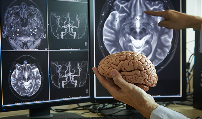
Understanding Your Brain Scan Results
Getting brain imaging done raises a question almost everyone asks: what does “normal” actually mean? And what does an abnormal finding mean for me?
A normal brain CT in an emergency setting is meaningful information. It says there’s no significant bleeding, no obvious large mass, no bone fracture. That’s not a small thing – that’s often exactly what needed to be ruled out in the moment. But a normal CT doesn’t confirm there are no small lesions, no early white matter changes, no subtle tumors.
A normal brain MRI carries more diagnostic weight. Given MRI’s superior sensitivity for soft tissue abnormalities, a normal result covers significantly more ground. It substantially reduces the likelihood of MS, significant brain tumor, major vascular malformation, or structural cause for many neurological symptoms.
Abnormal findings on either scan require context. An incidental finding – something seen on imaging that wasn’t the reason the scan was ordered – is surprisingly common. Small white matter changes, for example, appear on MRI in a notable percentage of adults over 50 and often have no clinical significance. These findings need interpretation by physicians who know your full clinical picture, not just the radiology report in isolation.
For a deeper look at what “normal” brain MRI results actually tell you – and what they don’t – our dedicated guide at what a normal brain MRI means walks through the details.
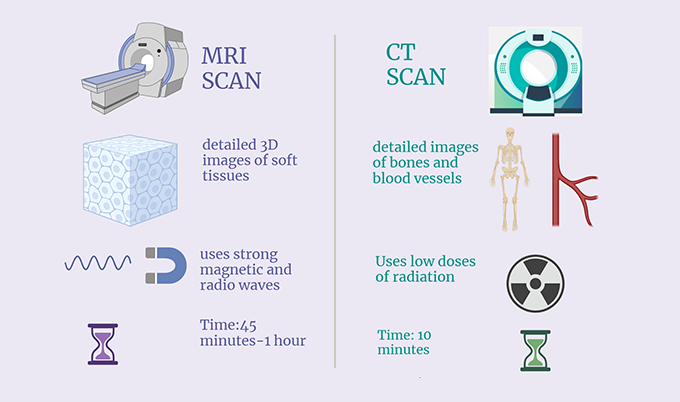
Brain MRI vs CT Scan: Side-by-Side Comparison
| Feature | Brain CT Scan | Brain MRI |
|---|---|---|
| Scan time | 5-10 minutes | 30-60 minutes |
| Radiation | Yes (low dose) | None |
| Best for emergencies | Yes | Less suited |
| Soft tissue detail | Moderate | Excellent |
| Bone detail | Excellent | Limited |
| Detects acute bleeding | Excellent | Good (varies by sequence) |
| Detects early stroke | Limited in first hours | Excellent (DWI sequence) |
| Detects MS lesions | Poor | Excellent |
| Tumor characterization | Moderate | Excellent |
| Metal implant limitations | Minimal | Significant |
| Cost | Generally lower | Generally higher |
The right scan is the one ordered for the right reason. Both technologies have genuine strengths – and genuine limitations. Neither replaces clinical judgment.
Who Should Consider Proactive Brain Scanning
Brain imaging isn’t only for emergencies or active neurological symptoms. There’s a growing conversation in preventive health about when proactive brain CT or MRI scanning makes sense – and what it can realistically find.
Certain groups have stronger reasons to consider proactive brain imaging:
- Individuals with a family history of brain aneurysm – First-degree relatives of patients with intracranial aneurysms have a meaningfully higher risk. MR angiography can screen for aneurysms before they cause symptoms.
- People with persistent or changing headache patterns – Headaches that shift in character, frequency, or severity – particularly those that wake someone from sleep or occur with exertion – warrant evaluation rather than assumption.
- Adults concerned about cognitive changes – Early memory changes, word-finding difficulties, or processing slowdowns that are noticed subjectively often prompt MRI evaluation to look for structural contributions.
- Those with cardiovascular risk factors – Hypertension, atrial fibrillation, and other vascular risk factors correlate with small vessel disease in the brain. Identifying this early allows for more aggressive risk factor management.
- Individuals with a history of significant head trauma – Prior TBI, even from years earlier, may have lasting structural implications that imaging can characterize.
The brain scan available through Craft Body Scan is designed for exactly this kind of proactive health monitoring – detecting conditions before they become crises. According to the American Heart Association, many serious brain and vascular conditions are silent until a major event occurs, which is precisely why screening in high-risk individuals matters.
✓ Should You Consider a Proactive Brain Scan?
- Family history of brain aneurysm, stroke, or intracranial tumors
- New or worsening headaches without a clear cause
- Unexplained memory changes, cognitive shifts, or word-finding problems
- History of significant head trauma or repeated concussions
- High blood pressure, atrial fibrillation, or other vascular risk factors
- Neurological symptoms (numbness, tingling, vision changes) with a normal CT result
Early detection changes outcomes. That’s true for heart disease, for cancer, and it’s equally true for neurological conditions. The brain is not an exception to the principle that finding problems before they cause permanent damage is better than waiting for symptoms to force the issue.
Is a Brain MRI or CT Scan Right for You?
These two imaging tools answer different questions. A brain CT scan asks: is there something urgent and structural happening right now – bleeding, fractures, large masses – that needs immediate attention? A brain MRI asks: what is the detailed architecture of this brain, and what subtle findings might explain what this patient is experiencing?
Knowing which question is being asked helps make sense of why one scan was chosen over the other. It also explains why a normal CT result doesn’t close every door, and why the same symptom might prompt different imaging choices depending on clinical context and timing.
If you’ve had a brain scan and want to understand what your results mean – or if you’re considering proactive brain imaging for the first time – the team at Craft Body Scan works with board-certified radiologists who review findings and communicate results clearly. Taking a proactive look at what’s happening in your brain is one of the more meaningful investments you can make in your long-term health.
Schedule your brain scan at Craft Body Scan today – because knowing what’s happening in your brain is always better than wondering.