Medical Disclaimer: This content is for educational purposes only and does not constitute medical advice, diagnosis, or treatment. Always consult with a qualified healthcare provider regarding any medical condition or treatment plan.
A normal brain MRI shows symmetrical brain structures, clear gray-white matter differentiation, normal-sized ventricles, and no signs of bleeding, swelling, tumors, or other acute abnormalities. Common report phrases like "unremarkable brain MRI" or "no acute intracranial abnormality" mean no urgent abnormal findings were seen.
- Symmetrical left and right hemispheres
- Normal ventricles and fluid spaces
- Clear gray matter and white matter boundaries
- No mass, bleed, stroke, swelling, or midline shift
If you've just received your brain MRI results, you're probably holding a report full of clinical language and wondering what it actually means for your health. That gap between receiving results and truly understanding them - that's where we come in. As radiologists who review hundreds of brain scans each month, we know how disorienting those black-and-white images can feel. The technical vocabulary creates distance when what you need is clarity.
Here's what we want you to know: "normal" on a brain MRI doesn't mean identical to every other brain. It means your scan shows no active disease, no structural problems requiring intervention, and no patterns that concern your radiologist. Understanding exactly what that means - and what it looks like - is what this guide covers.
What Does a Normal Brain MRI Look Like?
A normal brain MRI, when read by a board-certified radiologist, shows predictable patterns across multiple tissue types and anatomical regions. The brain's surface - the cerebral cortex - appears as a thin, consistent layer of gray matter wrapping around the outer edges. Beneath it, white matter forms the internal communication network, with characteristic signal intensity on each imaging sequence. Think of the cortex as your brain's command center and white matter as its fiber-optic cables.
Your ventricles appear as dark, fluid-filled spaces within the brain - symmetrical, appropriately sized, and proportional to the surrounding brain tissue. Cerebrospinal fluid cushions the brain and circulates through these spaces, removing metabolic waste in what researchers have described as the brain's glymphatic cleaning system. When ventricle size and shape fall within normal limits, that's one of the clearest signals a healthy brain MRI can show.
The 4 signs radiologists look for in a normal scan
Every normal brain MRI shares the same four foundational characteristics. When all four are present, a radiologist can report the scan as normal with confidence:
- Normal signal intensity across brain tissue - Gray matter, white matter, and cerebrospinal fluid each produce predictable signals on T1, T2, and FLAIR sequences. No focal areas of abnormal brightness or darkness appear across multiple sequences.
- Symmetrical hemispheric structure - Left and right brain hemispheres show balanced proportions and architecture, with no significant asymmetry beyond naturally occurring normal variation.
- No mass effect or midline shift - Brain structures remain centered. No lesions, collections, or swelling are pushing on surrounding tissue or displacing the midline.
- Normal vascular patterns - Major arteries and veins follow expected pathways. No evidence of acute stroke, aneurysm, or vascular malformation is present.
What "unremarkable" and "no acute intracranial abnormality" mean
Two phrases appear on normal brain MRI reports more than any others - and both confuse patients who expect plain language.
"Unremarkable brain MRI" is not dismissive. In radiology, "unremarkable" is a precise term meaning the radiologist found nothing worth noting - no abnormalities, no concerning patterns. It is one of the best sentences you can read in your report.
"No acute intracranial abnormality" is similarly reassuring. "Acute" refers to new or active findings - stroke, hemorrhage, herniation, or acute inflammation. The absence of acute findings means there is no emergency, no urgent intervention required, and no active disease process visible at the time of the scan.
Both phrases confirm the same thing: your brain MRI results fall within normal limits.
Symmetrical Structure
Left and right brain hemispheres show balanced proportions with no significant shifts or distortions.
Clear Gray/White Matter
Distinct boundaries between gray matter (nerve cells) and white matter (connecting fibers) throughout.
Normal Ventricles
Fluid-filled spaces appear appropriately sized and symmetrical - a sign of healthy cerebrospinal fluid circulation.
What Does a Brain MRI Show?
Understanding what a brain MRI can and cannot show helps put your results in context. MRI uses magnetic fields and radio waves - no radiation is involved. That combination produces soft-tissue contrast far superior to CT scanning, making it the preferred imaging tool for evaluating brain structure in detail.
What a brain MRI can detect
A brain MRI gives physicians a detailed view of brain tissue, fluid spaces, and blood vessel patterns. It can identify or rule out a wide range of conditions, including:
- Stroke and TIA (transient ischemic attack) - Diffusion-weighted imaging (DWI) sequences detect acute ischemic stroke within minutes to hours of onset.
- Brain tumors - Primary brain tumors and metastatic lesions appear as masses with characteristic signal patterns, often enhanced by contrast.
- Multiple sclerosis and demyelinating disease - White matter lesions in characteristic periventricular and juxtacortical locations signal MS on FLAIR sequences.
- Brain aneurysms and vascular malformations - MRA (magnetic resonance angiography) sequences visualize blood vessel abnormalities without catheter-based procedures.
- Intracranial hemorrhage - Bleeding at various stages appears with characteristic signal patterns on T1, T2, and gradient echo sequences.
- Hydrocephalus - Abnormal enlargement of the ventricular system indicates abnormal cerebrospinal fluid circulation.
- Encephalitis and brain abscess - Inflammation and infection produce distinct patterns of abnormal signal and enhancement.
- Structural abnormalities - Conditions like Chiari malformation, cortical dysplasia, or hippocampal atrophy are clearly visible.
What a brain MRI does not reliably show: early functional changes like mild cognitive decline, psychiatric conditions, or electrical activity (EEG measures that). MRI images structure - not function - unless specialized sequences like fMRI are used.
What a brain MRI with contrast vs without contrast can show
Most brain MRI screenings are performed without contrast as the first step. A standard non-contrast brain MRI covers the full range of structural evaluation - identifying stroke patterns, white matter changes, masses, and anatomical abnormalities with high accuracy.
Gadolinium contrast is added when the referring physician or radiologist needs to evaluate findings that only become visible when the blood-brain barrier is disrupted. According to the Radiological Society of North America, contrast-enhanced MRI is particularly valuable for:
- Detecting active tumor enhancement and distinguishing recurrent tumor from treatment effects
- Identifying areas of active inflammation or demyelination
- Evaluating suspected brain infection or abscess
- Assessing leptomeningeal disease (cancer spread to brain membranes)
If your report notes that contrast was administered, look for the phrase "no abnormal enhancement" in the findings section. That phrase confirms no blood-brain barrier breakdown was detected - a normal contrast MRI result. If you have questions about gadolinium contrast safety and alternatives, that guide covers the evidence in full.
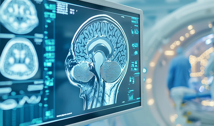
Normal Brain MRI Images and Anatomy

When radiologists describe what a healthy brain MRI looks like, they're reviewing multiple anatomical structures across several imaging planes. Each structure has a predictable, normal appearance - and recognizing that pattern is exactly what board-certified radiologists are trained to do.
Gray matter vs white matter
The brain's two primary tissue types produce distinct signals on MRI. Understanding this difference helps decode your report language.
Gray matter consists primarily of nerve cell bodies - the neurons that process information. On T1-weighted images, gray matter appears slightly darker than white matter. It forms the cerebral cortex along the brain's outer surface, and also makes up deep structures including the basal ganglia, thalamus, and hippocampi. In a healthy brain MRI, gray matter shows consistent thickness across the cortex with no focal areas of thinning or abnormal signal.
White matter consists of myelinated nerve fibers that carry signals between brain regions - the brain's internal communication infrastructure. It appears lighter than gray matter on T1 sequences. A normal brain MRI shows white matter with uniform signal intensity and a clear, predictable distribution. When white matter shows small areas of increased signal on T2 or FLAIR sequences - called white matter hyperintensities - the significance depends heavily on age, number, location, and pattern.
Small, scattered white matter hyperintensities in older adults often represent normal vascular aging and require no intervention. When they become extensive, form specific patterns, or appear in younger patients, they may indicate conditions like chronic microvascular ischemic disease or demyelinating conditions. Your radiologist's report will classify them with clinical context.
Ventricles, cerebellum, and brainstem
Three additional structures receive close attention on every normal brain MRI read:
Ventricles: Four interconnected chambers - two lateral ventricles, the third ventricle, and the fourth ventricle - hold and circulate cerebrospinal fluid. Normal ventricles appear dark on T1-weighted images, bright on T2 images, and dark on FLAIR sequences (the FLAIR sequence suppresses CSF signal specifically). Their size should be proportional to the surrounding brain volume. The ventricles naturally enlarge mildly with age as brain volume decreases - this is expected and documented in the report as age-appropriate.
Cerebellum: Located at the base of the brain, the cerebellum controls coordination, balance, and fine motor function. Its distinctive folded appearance - smaller and more tightly packed than the cerebral cortex - should show clear definition between folia (the individual folds) and preserved gray-white differentiation. Any asymmetry, atrophy, or abnormal signal here warrants attention.
Brainstem: The connection between brain and spinal cord shows three clear segments - the midbrain, pons, and medulla - in a normal brain MRI. Each should show normal, homogeneous signal with preserved internal structure. The brainstem houses critical centers for breathing, heart rate, and consciousness, making its evaluation an essential part of every brain scan review.
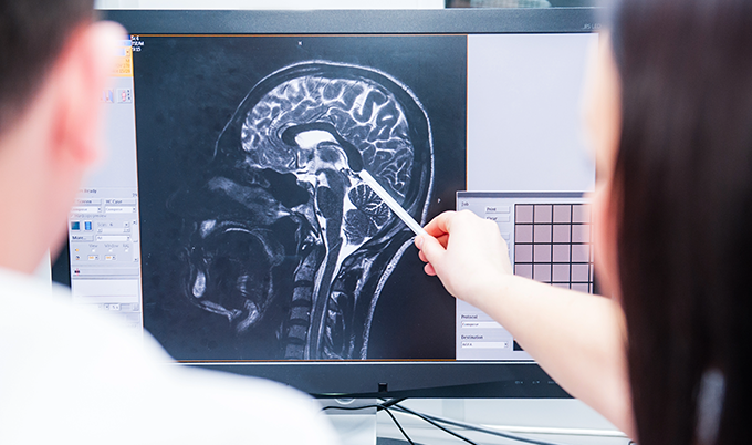
Axial, sagittal, and coronal brain MRI views
Radiologists review brain MRI images in three anatomical planes. Each plane reveals different structures and relationships - and together, they form the complete picture of a healthy brain MRI.
- Axial (transverse) view - Horizontal cross-sections from top to bottom, like slices through a loaf of bread. This is the view most people recognize from medical imaging on television. Axial slices clearly show the cerebral hemispheres, lateral ventricles, basal ganglia, and thalami. Most white matter assessment happens on axial views.
- Sagittal view - Slices from left to right through the midline, like cutting a book down the spine. The sagittal midline view is best for evaluating the corpus callosum, cerebellum, brainstem, and pituitary gland. It's the view radiologists use to confirm midline alignment and evaluate structures along the brain's central axis.
- Coronal view - Front-to-back slices that resemble a face-on photograph. Coronal images excel at evaluating the temporal lobes and hippocampal formations - structures critically important for memory - as well as the orbits and skull base.
A normal brain MRI shows consistent, expected signal patterns across all three planes with no focal abnormalities appearing in the same location on multiple sequences.
Normal vs Abnormal Brain MRI
A normal brain MRI is defined as much by what is absent as by what is present. Radiologists learn to recognize the expected patterns of a healthy brain scan - and then look deliberately for anything that deviates from those patterns. The split below reflects exactly how we frame that comparison.
- Clear, predictable gray/white matter boundaries with consistent signal intensity
- No masses, cysts, or tissue collections pressing on surrounding structures
- No areas of restricted diffusion - no stroke patterns on DWI sequences
- No evidence of bleeding at any stage (fresh or old hemosiderin deposits)
- Scattered, small white matter spots only - age-appropriate vascular changes
- Ventricles proportional to surrounding brain volume
- No abnormal enhancement after contrast injection
- Focal bright or dark spots disrupting expected tissue signal patterns
- Mass lesions that occupy space, shift midline, or enhance with contrast
- Restricted diffusion on DWI - bright patch in a vascular territory (acute stroke)
- Bright or dark regions indicating fresh or old hemorrhage
- Large confluent white matter changes or periventricular rings (e.g., MS pattern)
- Abnormally enlarged ventricles with compressed brain tissue (hydrocephalus)
- Unexpected enhancement suggesting disrupted blood-brain barrier
How tumors, bleeding, swelling, and white matter changes look different
Each type of abnormality has a characteristic pattern on brain MRI images. Knowing what radiologists look for makes it easier to understand a report that mentions a specific finding:
Tumors typically appear as areas of abnormal signal with defined or irregular borders. Primary brain tumors often show surrounding edema - bright signal on T2/FLAIR from fluid in the surrounding tissue. Metastatic lesions frequently appear at the gray-white matter junction and nearly always enhance with contrast as the blood-brain barrier is disrupted. A normal brain MRI shows none of these patterns.
Acute bleeding (hemorrhage) has a complex appearance that changes over time. Fresh blood appears bright on T1 images within hours. Over days and weeks, the signal evolves as hemoglobin breaks down. Old blood products - hemosiderin - appear as dark areas ("blooming") on gradient echo or susceptibility-weighted sequences. No such patterns appear on a healthy brain scan.
Stroke and ischemia affect brain tissue by cutting off blood supply. On diffusion-weighted imaging (DWI), acute ischemia appears as a bright signal - restricted diffusion - corresponding to the affected vascular territory. This signal appears within minutes of stroke onset. A normal brain MRI shows no restricted diffusion anywhere.
White matter disease ranges from incidental age-related changes to significant pathology. The pattern matters: periventricular white matter changes in a classic "periventricular halo" distribution raise concern for demyelinating disease. Subcortical punctate lesions scattered through deep white matter in an older adult more often represent normal vascular aging. A radiologist's job is to distinguish between them using location, pattern, and clinical context.
When white spots may be benign vs worth follow-up
White matter hyperintensities are among the most common "findings" mentioned in brain MRI reports - and among the most misunderstood. Here's how radiologists frame the distinction:
- Likely benign (normal aging) - Few in number, small (under 3mm), located in deep white matter, no periventricular involvement, patient over 50. These appear in a large proportion of healthy adults and typically do not require follow-up imaging.
- Worth documenting and monitoring - Moderate number, slightly larger, some periventricular location, patient under 50 with vascular risk factors. Your physician may recommend follow-up imaging in 1-2 years to assess stability.
- Requiring further evaluation - Numerous, confluent or large, periventricular pattern, enhancing lesions, or new compared to prior scans. These patterns warrant prompt clinical correlation and potentially additional sequences or specialist referral.
The bottom line: finding white spots on a brain MRI report does not automatically mean something is wrong. Context determines meaning - and a radiologist who has reviewed your full clinical picture is best positioned to explain what your specific findings mean for you.
How to Read Brain MRI Results and Reports

Most patients receive a written radiology report alongside - or instead of - a direct conversation with a radiologist. These reports follow a consistent structure across all imaging facilities. Knowing how to read brain MRI results means knowing that structure before you open the document.
How radiologists write findings and impression sections
Every brain MRI report contains the same core sections, reviewed in order. When radiologists interpret your scan, they follow a systematic protocol for consistent, complete evaluation:
Image Quality Assessment
We verify scan clarity and check for any technical issues - motion artifacts, positioning problems, or equipment anomalies - that might affect interpretation before evaluating findings.
Systematic Brain Review
We examine each brain region in order - frontal lobes, temporal lobes, parietal regions, occipital lobes, cerebellum, and brainstem - comparing structures bilaterally for symmetry and normal appearance.
Age-Appropriate Analysis
We consider your age and medical history to distinguish normal aging changes from concerning findings. What might appear concerning in a 30-year-old could represent completely normal aging in a 70-year-old.
Comprehensive Report
We provide clear, detailed brain scan results with explanations and actionable next steps - incorporating all imaging findings alongside your clinical information for a complete picture.
Common phrases patients see in brain MRI reports
The Findings section documents each brain structure in order - normal findings and abnormal ones alike. The Impression section is where the radiologist's bottom-line conclusion appears. If your scan was normal, that Impression section will contain one of the phrases in the glossary below.
If your report contains terminology you don't recognize, your ordering physician is the right person to walk you through it. At Craft Body Scan, our radiologists write brain scan results with clarity in mind and are available to explain findings when needed.
Common Variations That Can Still Be Normal
One of the most important things to understand about reading a normal brain MRI is that "normal" encompasses significant natural variation between individuals. Perfect symmetry does not exist in the human brain. What might initially look concerning to an untrained eye often represents completely normal anatomical variation - and a radiologist's training is built around knowing the difference.
Age-related changes
The brain changes predictably throughout the lifespan. Understanding these changes helps patients - and their physicians - correctly interpret brain scan results that mention age-related findings.
- Mild brain volume loss begins gradually in our thirties and accelerates after 60. This produces widening of the sulci (the grooves between brain folds) and mild ventricular enlargement. Both are expected findings on a healthy brain MRI in an older adult.
- White matter hyperintensities increase in prevalence with age. According to published neuroimaging data, they are present in a significant proportion of adults over 60 who are neurologically normal. Small, scattered periventricular and subcortical white matter changes in an older adult are typically documented as age-appropriate.
- Pineal gland calcification is extremely common with age. The pineal gland - a small endocrine structure at the brain's center - appears bright and dense on certain MRI sequences due to calcium deposits. This is a normal finding with no clinical significance.
- Choroid plexus calcification within the lateral ventricles is similarly normal and increases with age.
- Mild hippocampal volume reduction can be age-appropriate in older adults. Radiologists compare hippocampal volume against age-matched norms before drawing any conclusions.
Incidental findings that do not always signal disease
Sometimes a normal brain MRI reveals incidental findings - structures that appear slightly unusual but carry no clinical significance and require no treatment. These are documented precisely because they were seen, not because they are dangerous.
- Arachnoid cysts are fluid-filled spaces between brain tissue layers. Present since development, they are found in approximately 1% of the general population according to published neuroimaging studies. Most are completely asymptomatic and require no intervention.
- Developmental venous anomalies (DVAs) are variant venous drainage patterns. They appear as a caput medusae pattern on contrast-enhanced sequences and are considered normal anatomical variants. They do not require treatment.
- Prominent perivascular spaces (Virchow-Robin spaces) are enlarged fluid spaces along small blood vessels. In younger adults, they can appear quite prominent - particularly in the basal ganglia - without any pathological significance.
- Asymmetric ventricles are common. The occipital horns of the lateral ventricles in particular often differ in size between hemispheres. This falls within established normal ranges.
- Mega cisterna magna is an enlarged posterior fossa fluid space. Usually an incidental normal variant, it is documented in reports but rarely requires follow-up.
Having an incidental finding in your brain scan results does not mean something is wrong. It means the radiologist found something worth noting - and noted it. Your ordering physician can clarify whether any documented incidental findings need follow-up.
When to Get a Brain MRI
Symptoms that often lead to brain imaging
Proactive brain imaging serves two distinct purposes: evaluating symptoms and establishing a healthy baseline. Certain symptom patterns commonly lead physicians to order brain MRI as part of evaluation:
- New or changing headache patterns - particularly headaches that are severe, sudden in onset ("thunderclap"), change in character from prior headaches, or worsen with position or activity
- Neurological symptoms - numbness, weakness, vision changes, speech difficulty, coordination problems, or any focal neurological deficit
- Memory and cognitive concerns - especially when changes are noted by the patient or family and affect daily functioning
- First seizure - MRI is part of the standard evaluation for new-onset seizure to identify structural causes
- Family history of brain aneurysm or hereditary neurological conditions - such as CADASIL, familial aneurysm syndrome, or hereditary tumor syndromes
- Post-traumatic evaluation - following significant head injury when CT has not fully characterized the injury pattern
Contact sport athletes benefit from baseline brain imaging. When a future concussion or head injury occurs, having a prior normal brain MRI gives physicians a direct comparison point - an objective reference for detecting any changes that may have developed.
For those seeking preventive screening, a brain MRI scan can provide baseline documentation of healthy brain structure. Combined with a full body scan, this approach extends proactive health monitoring across the body's key organ systems. If you experience any anxiety about enclosed spaces, the guide to claustrophobia and MRI covers the full range of options for comfortable scanning.
When CT is used instead of MRI
MRI and CT (computed tomography) serve overlapping but distinct purposes in brain imaging. Understanding the difference helps patients ask better questions about why a specific test was ordered.
CT is the first choice in acute emergency settings - particularly when acute hemorrhage (bleeding) needs to be ruled out quickly. CT scans are faster than MRI, making them preferred when time matters most, such as in a stroke protocol. According to the American College of Radiology Appropriateness Criteria, CT is also preferred when MRI is contraindicated - in patients with certain implanted metal devices, pacemakers, or severe claustrophobia where sedation isn't available.
MRI is preferred for:
- Evaluating soft tissue brain structure in detail
- Non-emergency neurological evaluation
- White matter and demyelinating disease assessment
- Posterior fossa (brainstem and cerebellum) evaluation, where CT shows more artifact
- Any case where radiation exposure is a concern
If you're preparing for either type of scan, the guide to what to eat before a CT scan or MRI covers preparation guidelines for both.
Next Steps After Normal Brain MRI Results

Normal brain scan results are worth more than just relief in the moment. They represent a baseline - a documented record of your brain's healthy structure that becomes a reference point for every clinical decision made about your neurological health going forward.
Brain Health Actions After a Normal MRI
- Keep copies of your brain MRI results and radiologist's report - they're valuable for any future healthcare provider.
- Ask your physician about appropriate follow-up imaging intervals given your age, risk factors, and any incidental findings noted.
- Start or continue cardiovascular exercise - it directly improves cerebral blood flow and supports long-term brain health.
- Follow a diet rich in omega-3 fatty acids and antioxidants, both of which are associated with reduced neuroinflammation.
- Use your baseline results as context if new neurological symptoms develop - this can speed up future diagnostic processes significantly.
- Prioritize consistent sleep, as the brain's glymphatic system clears metabolic waste primarily during sleep.
Continued monitoring: Depending on your risk factors and any documented incidental findings, your physician may recommend follow-up imaging at specific intervals. Some conditions develop gradually, making periodic reassessment valuable for early detection. A repeat healthy brain MRI scan two to five years later can confirm stability of any documented variants.
Symptom awareness: Having a documented normal baseline means any future neurological symptoms can be compared against a known healthy state. If new concerns arise - whether headaches, memory changes, or coordination issues - that prior normal brain MRI provides immediate context and can actually accelerate the diagnostic process.
Documentation and records: Keep copies of your normal brain MRI report and images. These are valuable for any future healthcare provider you see - whether that's a neurologist, primary care physician, or specialist. Having your brain scan results on hand prevents unnecessary repeat imaging when you change providers or move to a new area.
Normal brain MRI results represent more than the absence of disease. They confirm that your brain shows healthy structure and function - a genuine foundation for informed neurological health decisions going forward.
Our board-certified radiologists at Craft Body Scan review brain MRI scans with advanced imaging technology and deliver clear, complete brain scan results - so you leave with answers, not more questions about your neurological wellness.
Schedule Your Brain Scan TodayContact us to learn more about our brain imaging services and how we support your proactive approach to health.