Medical Disclaimer: This article is for educational purposes only and does not constitute medical advice. Consult a qualified healthcare provider for any health concerns or before making decisions about medical imaging.
Most ovarian cysts are found by accident. A woman comes in for a routine pelvic exam, or a scan ordered for something unrelated picks up a fluid-filled structure on the ovary that was never causing symptoms in the first place. That is how common they are — the majority of women will develop at least one ovarian cyst during their reproductive years, and most resolve on their own within a few menstrual cycles without treatment.
But some do not resolve. Some grow large enough to cause real symptoms. Some turn out to be something more than a simple functional cyst. And when that happens, ovarian cyst imaging becomes the deciding factor in what comes next — whether that means watchful waiting, medication, or surgery.
Here is a clear breakdown of the symptoms that typically lead to imaging, and which tests identify ovarian cysts fastest and most accurately.
Symptoms That Usually Trigger Ovarian Cyst Imaging

Ovarian cysts occupy a wide spectrum. At one end are functional cysts — small, fluid-filled sacs that form as a normal part of the menstrual cycle and disappear without intervention. At the other end are complex masses with solid components, thick walls, or internal blood flow that warrant much closer attention. The symptoms a woman experiences often reflect where on that spectrum her cyst sits.
Persistent heaviness or bloated sensation in the lower abdomen, often on one side. Appears when a cyst grows large enough to press against surrounding structures.
EvaluateSharp or dull pain in the lower abdomen or pelvis, sometimes radiating to the lower back or thigh. Intermittent pain may indicate the cyst is shifting or twisting.
EvaluateDeep pelvic pain during or after sex is one of the more common reported symptoms and often prompts gynecological evaluation for cyst activity.
EvaluateCycles that change in length, flow, or cramping intensity without a clear explanation can sometimes be traced to cystic ovarian activity.
MonitorThe ER symptom. Sudden, sharp pain — sometimes with nausea, vomiting, or fever — can signal a ruptured cyst or ovarian torsion. Requires immediate care.
Seek ER ImmediatelyA large cyst pressing on the bladder can cause increased urgency to urinate or difficulty fully emptying the bladder.
MonitorWhen to Go to the Emergency Room
- Sudden, severe abdominal or pelvic pain that comes on rapidly and is unlike your normal discomfort
- Pain accompanied by fever and vomiting — a possible sign of ovarian torsion or a ruptured cyst with infection
- Lightheadedness or fainting alongside abdominal pain, which may indicate internal bleeding from a ruptured cyst
These symptoms require urgent evaluation — ovarian torsion can cut off blood supply to the ovary within hours. Do not wait for a scheduled appointment.
It is worth noting that many ovarian cysts cause no symptoms at all. A completely asymptomatic cyst found incidentally on imaging is not automatically cause for alarm — but it does need to be characterized, which is exactly what the imaging process is designed to do.
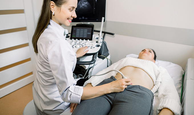
Transvaginal Ultrasound: The Fastest First Step
When a doctor suspects an ovarian cyst, the first imaging test ordered is almost always a pelvic ultrasound — specifically a transvaginal ultrasound (TVUS). This is considered the gold standard for initial ovarian cyst evaluation, and for good reason.
The procedure involves inserting a small probe into the vaginal canal. Because the probe sits directly adjacent to the ovaries, it produces high-resolution images without the interference of abdominal tissue, bowel gas, or body size. The exam takes roughly 20 to 30 minutes, uses no radiation, and is typically available same-day or within a day or two in most clinical settings.
What transvaginal ultrasound can do extremely well is distinguish simple cysts from complex ones. A simple cyst — round, thin-walled, filled with clear fluid, no internal structures — carries a very low risk of malignancy and usually requires only monitoring. A complex cyst, by contrast, may have internal debris, thick septations dividing the interior into compartments, solid components, or visible blood flow on color Doppler imaging. Each of those features changes the clinical picture significantly.
Research published in BJC Reports found ultrasound subjective assessment achieved sensitivity as high as 96.7% for distinguishing benign from malignant ovarian masses. That is a strong performance for a noninvasive test that requires no preparation and no contrast agent.
Transabdominal ultrasound — where the probe moves across the outside of the abdomen — is sometimes performed alongside transvaginal ultrasound to assess larger masses that extend beyond the pelvis. Both approaches are often done together as a complete pelvic ultrasound study.
What Ultrasound Can Tell You About a Cyst’s Type
Ultrasound does more than confirm that a cyst exists. It helps characterize what kind of cyst is present, which directly guides the next decision. Radiologists look for specific features that correspond to different cyst types.
When MRI Steps In
If transvaginal ultrasound identifies a cyst but cannot definitively characterize it — or if the findings are complex enough to require a more detailed look before a treatment decision — MRI is the natural next step.
MRI uses no radiation and provides superior soft tissue contrast compared to both ultrasound and CT. It is particularly useful for identifying the internal composition of a cyst: whether it contains blood, fat, mucin, or solid tissue. That distinction matters because it separates the vast majority of benign cysts from the minority that warrant surgical evaluation.
MRI has demonstrated sensitivity of 96% and specificity of 91% for distinguishing malignant ovarian tumors from benign ones. In cases where ultrasound returns an indeterminate result, MRI provides the additional clarity needed to avoid unnecessary surgeries while still catching the findings that require them.
The procedure involves lying inside an MRI scanner for 30 to 60 minutes. Multiple imaging sequences are used — T1-weighted images highlight fat and blood products, T2-weighted images highlight fluid and soft tissue detail, and diffusion-weighted imaging can help assess the nature of solid components. Patients are sometimes asked to fast for a few hours beforehand and may receive an antispasmodic medication to reduce bowel movement during imaging.
Whole-Body MRI and Incidental Ovarian Findings
For women who want comprehensive pelvic and abdominal evaluation without requiring a separate specialist referral for each organ system, Craft Body Scan's Whole-Body MRI Scan provides full pelvic imaging reviewed by board-certified radiologists — giving patients a broader picture of pelvic and abdominal health. Ovarian findings are regularly identified this way, often before symptoms develop, leaving far more clinical options available.
The Role of CT in Ovarian Cyst Evaluation
CT is not typically the first-choice imaging modality for ovarian cysts. It uses ionizing radiation, provides less soft tissue detail than MRI, and is generally less effective than ultrasound for characterizing cyst contents in the initial evaluation.
Where CT does play an important role is in emergency settings and in staging. When a woman presents to an emergency department with sudden severe abdominal pain, a CT scan of the abdomen and pelvis is often the fastest way to assess whether a cyst has ruptured, whether there is free fluid in the abdomen, and whether ovarian torsion — twisting of the ovary around its ligament, which cuts off blood supply — is a possibility. In those acute scenarios, speed matters more than soft tissue detail, and CT delivers fast, wide-field imaging that covers the entire abdomen and pelvis in a single pass.
Once a malignancy is confirmed, CT becomes central to staging — mapping how far a cancer has spread to lymph nodes, the peritoneum, or distant organs. That information directly determines treatment planning. But for the initial question of “what is this cyst and should I be concerned,” CT comes after ultrasound and MRI in the imaging sequence. See what a full body CT scan captures across all major organs.
The Imaging Sequence in Practice
Understanding how these tests typically flow helps clarify what to expect if you are referred for ovarian cyst imaging.
Ovarian Cyst Imaging — How Scans Are Ordered
First-Line Imaging
Characterizes cyst size and features. Assigns a risk category. Simple cysts with reassuring features move to monitoring with a repeat scan in 6–12 weeks.
Complex or indeterminate findings move to Step 2.
Indeterminate Findings
Provides detailed tissue characterization — blood, fat, mucin, solid tissue — to clarify what ultrasound could not definitively answer.
Helps avoid unnecessary surgeries while catching what matters.
Emergency or Staging
Used when speed is critical in an acute presentation, or when a confirmed malignancy needs to be staged before treatment begins.
Most women never reach this step.
| Imaging Modality | Sensitivity / Performance | Best For | When It's Used | Key Limitation |
|---|---|---|---|---|
| Transvaginal Ultrasound | 96.7% (benign vs. malignant) | First-line evaluation; simple vs. complex classification | First test, almost always | May not fully characterize complex or indeterminate masses |
| MRI | 96% sensitivity, 91% specificity | Tissue characterization; indeterminate ultrasound findings | After inconclusive ultrasound | Higher cost; longer exam time; limited access in some areas |
| CT Scan | Moderate; best for calcified or large masses | Emergency assessment; cancer staging | ER presentations; confirmed malignancy | Ionizing radiation; limited soft tissue detail vs. MRI |
| Transabdominal Ultrasound | Variable; complements TVUS | Larger masses extending beyond the pelvis | Often combined with TVUS in a full study | Less detail than transvaginal; affected by body habitus |
Taking Symptoms Seriously Matters
The frustrating reality of ovarian cysts is that the symptoms — pelvic pressure, irregular cycles, pain during sex — overlap with a long list of other gynecological conditions. That overlap means cysts are sometimes attributed to other causes and go unimaged for longer than they should be.
If you have persistent pelvic symptoms that have not been explained, or if a prior scan flagged an ovarian finding that has not been followed up, that is worth addressing. Early characterization of an ovarian mass — when it is small, when it has not yet shown signs of growth or complexity — gives doctors and patients far more options than waiting until symptoms become impossible to ignore.
Questions to Ask After an Ovarian Cyst Finding
- Is this a simple cyst or does it have complex features?
- What size is it, and has that changed since any prior imaging?
- Which type of cyst does this appear to be based on its characteristics?
- Does this finding explain my current symptoms?
- Do I need an MRI for further tissue characterization?
- When and how often should I return for a follow-up scan?
- Are there any features that would prompt earlier re-evaluation?
At Craft Body Scan, our Whole-Body MRI Scan and Full Body CT Scan evaluate pelvic organs as part of a broader look at what is happening throughout the body — with results reviewed by board-certified radiologists and detailed reports that give you something concrete to discuss with your doctor. No referral needed. Transparent pricing. Real answers.
Schedule your Craft Body Scan today and take the first step toward knowing what is actually going on inside your body.