When shooting pain radiates down your leg, doctors order a lumbar MRI for sciatica to pinpoint exactly what’s compressing your nerve. As someone who specializes in diagnostic imaging, I’ve worked with thousands of patients who finally got answers after months of unexplained leg pain.
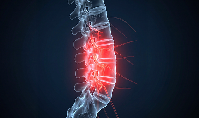
A lumbar MRI for sciatica uses powerful magnetic fields to create detailed cross-sectional images of your lower spine. These images reveal herniated discs, bone spurs, and inflammation pressing against nerve roots.
This painless 30-45 minute scan shows precisely which nerve is compressed and why. That information determines whether you need physical therapy, injections, or surgery.
Understanding Sciatica and Why Lumbar MRI for Sciatica Matters
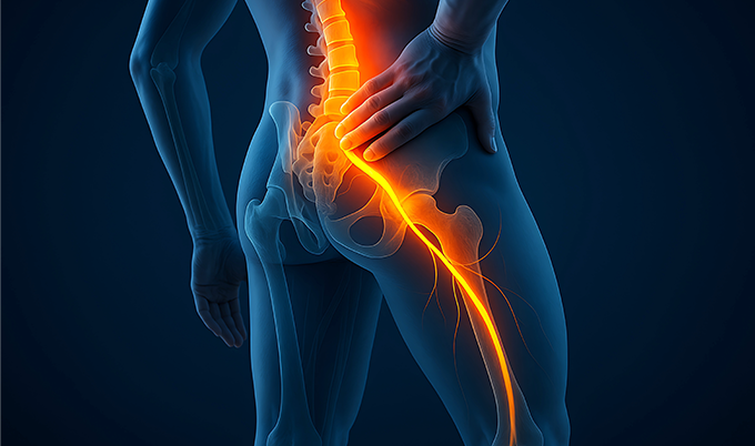
Sciatica affects 40% of adults at some point in their lives. The sharp, burning pain travels from your lower back through your buttock and down one leg.
Some patients describe electric shocks. Others report constant aching with numbness.
The sciatic nerve forms from five nerve roots that exit your lumbar spine. When disc material compresses these roots, pain radiates along the entire nerve pathway.
Clinical examination reveals which nerve is affected. Your doctor tests reflexes, muscle strength, and sensation patterns.
But physical exams can’t show what’s causing the compression. That’s where lumbar MRI for sciatica becomes essential for accurate diagnosis.
 Why Doctors Order Lumbar MRI for Sciatica
- Reveals soft tissues invisible on X-rays (discs, nerves, ligaments)
- Pinpoints compression location with millimeter precision
- Guides treatment decisions between conservative care and surgery
- Excludes red flag conditions requiring emergency intervention
- Plans injection placement for epidural steroid procedures
- Documents baseline severity for tracking progression
- Predicts treatment success using contrast enhancement patterns
How Lumbar MRI for Sciatica Detects Nerve Root Compression
The physics behind lumbar MRI for sciatica exploits hydrogen atoms in your body’s water molecules. When you enter the powerful magnetic field (1.5 or 3 Tesla), hydrogen protons align with that field.
Radiofrequency pulses knock these protons out of alignment. As they snap back, they emit signals your body creates.
The scanner detects these signals and computers convert them into detailed cross-sectional images. Different tissues emit distinct signals on lumbar MRI for sciatica scans.
Herniated disc material appears bright on T2-weighted images. Nerve roots show intermediate signal. Bone appears dark due to low water content.
These signal differences reveal compression patterns doctors use to guide treatment. The scanning sequences capture multiple views taking 3-5 minutes each.
Multiple Imaging Sequences on Lumbar MRI for Sciatica
Lumbar MRI for sciatica uses specialized sequences to highlight specific problems. T1-weighted images show anatomical detail and help identify fat tissue.
T2-weighted images highlight fluid and inflammation around compressed nerves. STIR sequences reveal bone marrow edema indicating recent injury or stress reactions.
Radiologists examine hundreds of image slices from multiple angles. Sagittal views show disc alignment and herniation extent on lumbar MRI for sciatica.
Axial views reveal nerve root compression severity. Coronal views detect lateral abnormalities affecting nerve exit holes.
| MRI Sequence | What It Shows | Clinical Value for Sciatica |
|---|---|---|
| T1-Weighted | Anatomical detail, fat tissue | Identifies nerve root location |
| T2-Weighted | Fluid, disc hydration, pathology | Highlights herniated discs and nerve edema |
| STIR Sequence | Bone marrow edema, inflammation | Detects recent injury or stress reactions |
| Contrast-Enhanced | Blood flow, active inflammation | Predicts conservative treatment success |
What Lumbar MRI for Sciatica Reveals About Disc Herniations
Research shows 95% of lumbar disc herniations occur at L4-L5 or L5-S1. These lower levels carry the highest loads and experience the most movement stress.
Herniated discs appear as bulging material protruding beyond normal disc boundaries. The inner nucleus pulposus pushes through tears in the outer annulus fibrosus on lumbar MRI for sciatica images.
This creates the pressure that compresses nerve roots causing your symptoms. Lumbar MRI for sciatica classifies herniation severity using standardized terminology that guides treatment.
Bulge: The entire disc circumference extends beyond vertebral edges but the outer wall remains intact. These often improve with conservative care alone.
Protrusion: Disc material pushes through a focal area but remains connected to the main disc. This is the most common finding on lumbar MRI for sciatica.
Extrusion: Disc material breaks through the outer wall and separates from the disc. These cause more severe compression requiring intervention.
Sequestration: Disc fragments migrate away from the original herniation site. These often require surgical removal for symptom resolution.
Disc Degeneration Patterns on Lumbar MRI for Sciatica
The scan shows disc height loss indicating degeneration throughout your spine. Dark signal intensity on T2-weighted images reveals disc desiccation.
This is loss of water content that normally cushions vertebrae. Studies show disc degeneration affects 98% of patients with lumbar herniations.
This suggests degeneration occurs throughout the spine. It’s not just at symptomatic levels causing your current sciatica symptoms visible on lumbar MRI for sciatica.
Lumbar MRI for sciatica also detects Modic changes – signal abnormalities in vertebrae adjacent to degenerated discs. Type 1 changes show bone marrow edema correlating with active inflammation and pain intensity.
Type 2 changes show fatty infiltration indicating chronic degeneration. These findings help predict which patients respond better to specific treatments.
Nerve Root Compression Patterns Revealed by Lumbar MRI for Sciatica
Lumbar MRI for sciatica shows exactly which nerve is compressed and how severely compressed it appears. Radiologists grade compression using standardized systems that directly guide treatment decisions.
Grade 1 means the nerve root contacts disc material but maintains normal shape. This often improves with conservative treatment protocols.
Grade 2 means the nerve root is displaced or deformed by compression visible on lumbar MRI for sciatica. These patients typically need more aggressive intervention approaches.
Grade 3 means the nerve root is severely compressed and difficult to visualize on the scan. These cases often require surgical decompression for symptom relief.
Understanding Your Specific Nerve Compression Symptoms
Each lumbar nerve root produces distinct symptoms that correlate with anatomical findings. Lumbar MRI for sciatica helps match your symptoms to visible compression.
L4 nerve compression causes anterior thigh pain and medial leg numbness. You experience knee extension weakness making stair climbing difficult.
Patients struggle climbing stairs and notice decreased patellar reflex. Lumbar MRI for sciatica shows compression at the L3-L4 or L4-L5 disc level.
L5 nerve compression causes lateral leg pain traveling to the dorsum of your foot. You develop foot drop and big toe weakness visible during examination.
Walking on heels becomes difficult or impossible. This is the most common sciatica pattern seen on lumbar MRI for sciatica imaging.
S1 nerve compression causes posterior leg pain radiating to your lateral foot and heel. You experience ankle weakness and difficulty standing on toes during testing.
The Achilles reflex disappears completely. Lumbar MRI for sciatica typically shows compression at the L5-S1 disc level.
Lumbar MRI for sciatica reveals specific compression mechanisms determining optimal surgical approach. Posterolateral disc herniations compress the traversing nerve root traveling down to the next spinal level.
Far lateral herniations compress the exiting nerve root requiring different surgical access. This anatomical detail on lumbar MRI for sciatica is crucial for surgical planning.
The scan shows inflammatory changes surrounding compressed nerves throughout the spine. Increased signal intensity on T2-weighted images indicates nerve edema and swelling.
This inflammation visible on lumbar MRI for sciatica contributes to your pain intensity. Sometimes inflammation causes more pain than mechanical compression alone.
 What Your Symptoms Tell Doctors Before Lumbar MRI
- Anterior thigh pain suggests L4 nerve compression at L3-L4 or L4-L5
- Lateral leg pain with foot drop indicates L5 compression at L4-L5
- Posterior leg pain to heel points to S1 compression at L5-S1
- Bilateral leg symptoms raise concern for central canal stenosis
- Bowel/bladder changes require emergency MRI for cauda equina syndrome
- Progressive weakness indicates urgent imaging need regardless of pain level
How Lumbar MRI for Sciatica Guides Conservative Treatment Planning

Lumbar MRI for sciatica determines appropriate conservative treatments before considering surgical options. Physical therapists use imaging results to design safe exercise programs targeting your specific pathology.
Patients with anterior disc herniations avoid flexion exercises that increase forward pressure on nerves. Those with posterior herniations avoid extension exercises that worsen compression.
The scan guides these critical activity modifications protecting your nerves. Epidural steroid injections require precise needle placement for maximum effectiveness.
Lumbar MRI for sciatica shows exact compression locations for targeting these injections accurately. Interventional pain physicians inject specific nerve roots under fluoroscopic guidance using your MRI results.
The scan reveals which nerve root to inject for symptom relief. L5 nerve root for L4-L5 herniations or S1 nerve root for L5-S1 herniations shown on lumbar MRI for sciatica.
This precision improves injection success rates significantly compared to blind injection techniques.
Contrast-Enhanced Lumbar MRI for Sciatica Predicts Success
Contrast-enhanced lumbar MRI for sciatica uses gadolinium injection to reveal inflammation patterns around compressed nerves. This specialized imaging predicts which patients will improve without surgery based on enhancement patterns.
Rim enhancement – a bright ring surrounding herniated disc material – indicates active inflammatory response. Research shows 88% of herniations demonstrate this pattern on contrast studies using lumbar MRI for sciatica protocols.
Studies reveal patients with rim enhancement have 95.5% success rate with conservative treatment approaches. Those without enhancement have only 33.3% success rate with non-surgical management.
Two-thirds of patients without rim enhancement visible on lumbar MRI for sciatica ultimately require surgical intervention. The thickness of rim enhancement correlates with inflammation severity and healing potential.
Measurements range from 1-3.8mm across different patients scanned. Thicker enhancement suggests more robust healing response and better conservative treatment outcomes according to research from the National Library of Medicine.
This information helps doctors counsel patients about realistic treatment timelines. Presence of rim enhancement on lumbar MRI for sciatica supports continuing conservative care approaches.
Absence of enhancement suggests earlier surgical consultation may be appropriate for symptom relief.
Planning Surgical Interventions Using Lumbar MRI for Sciatica
When conservative treatment fails after 6-8 weeks, lumbar MRI for sciatica guides complete surgical planning. Surgeons need precise anatomical information visible on imaging to achieve optimal nerve decompression during procedures.
The scan determines surgical approach based on herniation location and type visible on lumbar MRI for sciatica:
Microdiscectomy for contained disc herniations: Surgeons remove protruding disc material through small incision targeting compression. Success rates exceed 90% when imaging shows clear nerve compression.
Laminectomy for spinal stenosis: Removing bone and ligament decompresses nerves effectively. Lumbar MRI for sciatica shows exactly how much tissue requires removal.
Foraminotomy for foraminal stenosis: Enlarging neural exit holes relieves compression at specific levels. The scan reveals which levels need decompression based on stenosis severity.
Fusion for instability: Stabilizing motion segments with hardware when multiple levels show degeneration. Used when lumbar MRI for sciatica demonstrates advanced degenerative changes.
How Lumbar MRI Findings Predict Surgical Outcomes
Research shows surgical outcomes correlate strongly with findings visible on lumbar MRI for sciatica. Patients with definite nerve root compression on imaging report 91% perceived recovery after surgery.
Those without clear compression on lumbar MRI for sciatica report only 50% recovery rates. This data emphasizes the importance of correlating symptoms with imaging findings before proceeding.
Sequestrated disc fragments that have migrated require careful surgical planning using detailed imaging. Lumbar MRI for sciatica shows fragment location – cranial or caudal migration from original herniation site.
Surgeons must locate these fragments during decompression procedures for complete symptom resolution. The imaging reveals anatomical variations affecting optimal surgical approach selection.
Conjoined nerve roots, transitional vertebrae, and vascular anomalies appear on detailed scans. Recognizing these variations on lumbar MRI for sciatica prevents surgical complications and improves patient outcomes.
When MRI Findings Point Toward Surgery
If your lumbar MRI shows moderate to severe nerve compression, or disc material that has broken through or separated from the disc entirely, physical therapy and medications probably aren’t going to fix the problem on their own. That’s when surgery enters the conversation. Most people hear “back surgery” and think of the traditional options: microdiscectomy, laminectomy, or spinal fusion. Those procedures have been around for a long time, and they can work, but they come with real downsides. Recovery takes weeks to months, complication rates are higher than most patients expect, and fusion permanently eliminates movement at the treated level of the spine.
What a lot of people don’t realize is that newer, less invasive options exist. Lumbar laser disc repair, for example, uses a tiny incision (smaller than a pencil eraser) and an endoscopic camera to remove the herniated disc material and inflamed tissue causing the nerve compression. No bone is cut. No muscle is severed. If your MRI clearly shows a disc pushing on a nerve root, it’s worth asking your spine specialist about the full range of procedures available to you, including laser-based options, before deciding on a treatment path.
Understanding Important Limitations of Lumbar MRI for Sciatica
Lumbar MRI for sciatica has important limitations requiring careful clinical correlation with your symptoms. Asymptomatic findings are extremely common in the general population without back pain.
Research shows 52% of pain-free people have disc bulges on imaging studies. Twenty-seven percent have protrusions visible on scans.
One percent have extrusions despite having no symptoms whatsoever. These incidental findings on lumbar MRI for sciatica shouldn’t trigger unnecessary treatment.
Studies reveal disc herniation presence doesn’t perfectly predict clinical outcomes. Patients can improve dramatically despite persistent herniation visible on follow-up imaging.
Conversely, patients with complete herniation resolution on lumbar MRI for sciatica may continue experiencing significant pain. The scan captures a single moment in time during dynamic disease processes.
Disc herniations change constantly – enlarging, shrinking, or resorbing spontaneously over weeks and months. Initial imaging may not represent current status when symptoms change.
Some nerve compression occurs outside the spine where standard protocols don’t image adequately. Piriformis syndrome, pelvic tumors, and extraspinal nerve entrapment produce sciatica-like symptoms.
Specialized imaging sequences beyond standard lumbar MRI for sciatica protocols may be needed. These conditions require dedicated pelvic or hip imaging for accurate diagnosis.
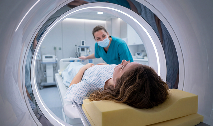
What to Expect During Your Lumbar MRI for Sciatica Examination
Preparing for lumbar MRI for sciatica requires removing all metal objects before entering the scanner room. Jewelry, watches, phones, keys, and clothing with metal fasteners interfere with magnetic fields.
Staff screens for implanted devices before proceeding with scanning procedures. Pacemakers, cochlear implants, and some orthopedic hardware may contraindicate MRI entirely.
Modern devices increasingly use MRI-compatible materials that allow safe scanning. You lie face-up on a narrow motorized table during lumbar MRI for sciatica.
Technicians position a spine coil around your lower back providing optimal image quality. They may place padding under your knees for comfort during examination.
The table slides into the scanner’s tunnel opening for imaging. Your head and upper body may remain outside the tunnel opening.
This open design reduces claustrophobia concerns for most patients undergoing lumbar MRI for sciatica. The actual scan requires absolute stillness throughout multiple sequences.
Each imaging sequence takes 3-5 minutes to complete properly. Any movement blurs images and necessitates sequence repeats extending exam time significantly.
The machine makes loud banging and buzzing noises during scanning. Most facilities provide earplugs or headphones playing music to reduce noise discomfort during lumbar MRI for sciatica.
If contrast is needed, technicians pause scanning midway through examination. They inject gadolinium through an IV line placed in your arm beforehand.
You may feel coolness spreading as the contrast circulates throughout your body. Scanning resumes to capture contrast-enhanced sequences showing inflammation patterns visible on lumbar MRI for sciatica.
Post-procedure recovery is immediate after completing the examination. You can resume normal activities unless you received sedation medication beforehand.
Gadolinium contrast exits through your kidneys within 24 hours completely.
Lumbar MRI for Sciatica Cost and Insurance Coverage
Lumbar MRI for sciatica costs $400-$6,500 without insurance depending on facility type selected. Independent imaging centers typically charge significantly less than hospital-based facilities for identical examinations.
With insurance coverage, out-of-pocket costs range from $50-$500 on average. This depends on your specific plan’s deductibles, copays, and coinsurance requirements.
Many insurers require prior authorization before approving lumbar MRI for sciatica requests. Insurance companies mandate conservative treatment documentation before approval.
You typically need 6 weeks of treatment including physical therapy before approval. This requirement ensures appropriate resource utilization and cost control.
Emergency presentations bypass authorization requirements when red flag symptoms exist requiring immediate imaging:
– Progressive weakness developing over hours or days
– Saddle anesthesia (numbness around rectum and genitals)
– Bowel or bladder dysfunction indicating cauda equina syndrome
– Fever accompanying back pain suggesting spinal infection
– History of cancer raising metastasis concerns
– Unexplained weight loss with back pain
– Age over 70 with new onset severe pain
Scheduling timeframes vary by urgency classification for lumbar MRI for sciatica. Emergent scans happen within hours when red flags exist.
Urgent scans occur within 24-72 hours for progressive symptoms requiring prompt evaluation. Routine scans may require waiting 2-4 weeks at busy imaging facilities.
Making Informed Treatment Decisions About Lumbar MRI for Sciatica
Understanding when lumbar MRI for sciatica adds clinical value helps you advocate for appropriate care. Acute sciatica lasting less than 6 weeks rarely requires imaging unless red flag symptoms exist.
Studies show over 85% of acute disc herniation symptoms resolve within 8-12 weeks. Conservative treatment alone achieves excellent outcomes without surgery.
Only 5-10% of sciatica patients ultimately require surgical intervention for symptom resolution. Persistent or progressive symptoms justify lumbar MRI for sciatica examination for diagnosis.
When leg pain continues despite 6 weeks of appropriate treatment, imaging provides diagnostic clarity. Lumbar MRI for sciatica reveals specific causes guiding treatment adjustments effectively.
Progressive neurological deficits warrant immediate imaging evaluation regardless of pain levels. Worsening weakness, expanding numbness, or new bowel/bladder symptoms require urgent assessment.
At Craft Body Scan, we understand the anxiety accompanying unexplained leg pain. Our board-certified radiologists provide detailed interpretations that help you make informed treatment decisions.
While we focus on preventive full body scanning, we recognize the critical importance of targeted diagnostic imaging. According to the American College of Radiology, MRI is the preferred imaging modality for evaluating lumbar spine pathology when radicular symptoms persist beyond initial conservative management.
The scan results inform shared decision-making between you and your medical team effectively. Your physician interprets lumbar MRI for sciatica findings alongside your symptoms and examination findings.
Imaging abnormalities don’t automatically mandate surgery in every single case presenting for evaluation. Some herniations improve spontaneously through natural resorption processes over time.
The body’s inflammatory response breaks down herniated disc material over several months. This natural healing occurs in more than 50% of symptomatic herniations documented on imaging.
Lumbar MRI for sciatica provides the essential roadmap for your personalized treatment journey. Whether that journey involves continued physical therapy, targeted injections, or surgical decompression depends on findings.
Understanding the scan’s capabilities and limitations empowers informed discussions with your medical team. Schedule your consultation to discuss whether lumbar spine imaging is appropriate for your specific symptoms.