You’re in the shower when your hand brushes against something unexpected––a hard lump under skin that wasn’t there before. Your mind immediately races to worst-case scenarios. Should you be concerned? The answer depends on specific characteristics that distinguish benign growths from those requiring immediate medical attention.
Take a breath. Instead of letting fear take over, let’s look at what clear medical guidelines can tell you about assessing the situation properly.
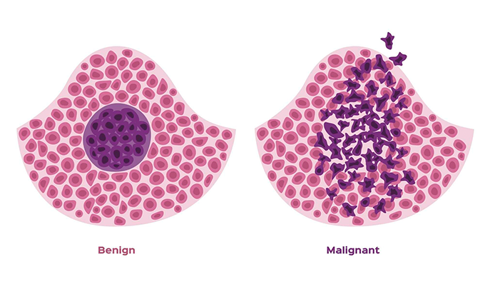
Here’s what the medical evidence shows: most hard lumps under skin are benign, meaning they’re not cancerous and don’t pose serious health risks. However, certain characteristics––including rapid growth, fixed attachment to surrounding tissues, and specific locations––do warrant prompt medical evaluation.
The key is understanding when a hard lump under skin represents normal tissue variations versus concerning changes. Benign lumps like cysts, lipomas, and dermatofibromas account for the vast majority of skin growths. These typically feel soft to firm, move easily when touched, and grow slowly over months or years.
In contrast, concerning hard lumps under skin often feel rock-hard, remain fixed when you try to move them, and may grow rapidly over days to weeks. Cancer specialists note that malignant lumps are typically large, hard, painless, and appear spontaneously without obvious cause.
Here’s the reality: while most skin lumps prove harmless, knowing which characteristics require immediate evaluation can give you peace of mind and ensure you get appropriate medical care when needed.
Understanding Hard Lumps Beyond Initial Fear

Hard lumps under skin form through various biological processes, most of which represent normal responses to injury, hormonal changes, or genetic predisposition. Understanding these mechanisms helps you distinguish between concerning and benign findings.
Your skin and underlying tissues are constantly undergoing cellular renewal, sometimes creating pockets where normal processes go a bit awry. Cysts develop when oil glands become blocked or hair follicles get clogged with dead skin cells, and inflammatory responses like granulomas can form as tissue reactions. These feel firm when deep beneath the surface but remain completely benign.
Hard lumps under skin follow predictable patterns based on their underlying cause. Benign hard lumps under skin typically feel smooth, well-defined, and mobile when gentle pressure is applied. Concerning hard lumps under skin often feel irregular, poorly defined, and fixed to deeper structures.
Location also influences significance. Certain areas of the body more commonly develop specific types of lumps. For instance, lipomas frequently appear on the back, arms, and shoulders, while dermatofibromas commonly develop on the legs and arms, particularly in areas prone to minor injuries.
The timeline of development provides crucial diagnostic clues. Benign growths typically develop gradually over months to years, allowing surrounding tissues to adapt. Rapid development over days to weeks may indicate more aggressive processes requiring immediate evaluation.
Most importantly, your body’s response to these lumps differs significantly between benign and concerning causes. Benign lumps rarely cause systemic symptoms, while problematic growths may be associated with unexplained weight loss, persistent fatigue, or other concerning symptoms.
Complete Guide to Hard Lumps Under Skin
This comprehensive breakdown covers the most common causes of hard lumps under skin, helping you understand what you’re dealing with and when medical evaluation becomes necessary.
Benign Causes of Hard Lumps
The majority of hard lumps under skin result from benign conditions that pose no serious health threat. Understanding these common causes provides reassurance and helps guide appropriate care decisions.
Epidermoid and sebaceous cysts represent the most common cause of hard lumps under skin. These develop when skin cells move deeper into tissue rather than shedding normally:
- Feel firm to hard depending on their depth beneath the skin surface
- Often have a small central opening (punctum) that may be difficult to see
- Can become inflamed if infected, causing redness, warmth, and tenderness
- Most commonly appear on the face, neck, chest, back, and genital areas
- Treatment involves complete removal of the cyst wall to prevent recurrence
Dermatofibromas are hard, brown or red lumps that develop in the dermis (deep skin layer):
- Feel very firm and well-attached to surrounding skin
- Usually develop on exposed areas like legs, arms, and back
- Often result from minor injuries like insect bites or small cuts
- May feel itchy or tender occasionally but usually cause no symptoms
- Remain stable in size once formed and don’t become cancerous
Lipomas typically feel soft but can feel firmer when they contain fibrous tissue or blood vessels:
- Most common in adults 40-60 years old and tend to run in families
- Feel soft and doughy in most cases but fibrolipomas feel firmer
- Move easily under the skin when gently pressed
- Rarely cause symptoms unless they involve nerves or blood vessels
- Treatment only needed if painful, growing rapidly, or cosmetically bothersome
Ganglion cysts are fluid-filled lumps that develop near joints:
- Feel firm and round and may change size with activity
- Most commonly appear on wrists, hands, and feet
- May grow larger with activity and shrink with rest
- Can be painful if they press on nearby nerves
- Often resolve spontaneously without treatment
Warning Signs That Require Medical Attention
While most hard lumps under skin are benign, certain characteristics indicate the need for prompt medical evaluation. Recognition of these warning signs ensures you get appropriate and timely care.
Physical characteristics of concerning lumps:
- Rock-hard texture that feels distinctly different from surrounding tissues
- Fixed attachment to deeper structures, not moving when you try to manipulate it
- Irregular borders or poorly defined edges rather than smooth, round contours
- Rapid growth over days to weeks rather than months to years
- Large size – lumps larger than 2 inches (about the size of a golf ball) warrant evaluation
Associated symptoms that increase concern:
- Skin changes over the lump including redness, ulceration, or unusual coloring
- Bleeding or discharge from the lump or surrounding skin
- Pain or tenderness that develops without obvious injury or infection
- Numbness or tingling in the area around the lump
- Systemic symptoms like unexplained weight loss, fever, or persistent fatigue
Timeline factors requiring attention:
- New lumps appearing after age 40 should be evaluated more promptly
- Changes in existing lumps including altered size, shape, or texture
- Lumps persisting beyond 2-4 weeks without signs of resolution
- Multiple new lumps appearing simultaneously in different body areas
Location-Specific Considerations

The location where a hard lump under skin develops influences both its likely cause and the urgency of evaluation. Certain body areas require more immediate attention due to their proximity to vital structures.
Head and neck lumps require particular attention due to critical anatomical structures:
- Scalp lumps may represent cysts, lipomas, or concerning growths requiring evaluation
- Neck lumps could indicate swollen lymph nodes from infection or malignancy
- Behind the ear – lumps in this area often represent lymph nodes responding to local infections
- Jaw area – may indicate dental infections, salivary gland problems, or lymph node issues
Torso lumps present different considerations based on specific location:
- Chest wall – may represent lipomas, cysts, or breast tissue changes requiring evaluation
- Back lumps – commonly lipomas or cysts, but rapid growth warrants assessment
- Abdominal wall – could indicate hernias, lipomas, or deeper abdominal issues
- Groin area – may represent swollen lymph nodes, hernias, or other conditions
Extremity lumps often relate to activity and local factors:
- Arms and shoulders – frequently lipomas, cysts, or activity-related swellings
- Hands and wrists – commonly ganglion cysts or tendon-related swellings
- Legs and thighs – often dermatofibromas, lipomas, or injury-related lumps
- Feet and ankles – may indicate ganglion cysts, bursitis, or other local conditions
When Hard Lumps Indicate Cancer
While rare, some hard lumps under skin can indicate malignant conditions. Understanding cancer-related characteristics helps ensure appropriate and timely evaluation when warranted through comprehensive cancer screening.
Soft tissue sarcomas represent the primary concern for hard lumps in soft tissues:
- Feel very hard and are typically painless in early stages
- Grow steadily over weeks to months, becoming noticeably larger
- Most commonly develop in legs, arms, chest, or behind the abdomen
- May reach significant size before causing symptoms
- Account for less than 1% of all adult cancers but require prompt attention
Metastatic deposits can appear as hard lumps when cancer spreads from elsewhere:
- Often multiple lumps appearing in different locations
- May be associated with known cancer history
- Can appear rapidly and grow quickly
- Often accompanied by other symptoms of cancer progression
Lymphoma can present as hard, enlarged lymph nodes under the skin:
- Feel firm and rubbery rather than soft like infected nodes
- Often painless and may appear in multiple locations
- May be accompanied by fever, night sweats, and weight loss
- Persist and grow rather than shrinking like infection-related swelling
Skin cancers including basal cell and squamous cell carcinoma can present as hard lumps:
- Usually develop on sun-exposed areas of skin
- May ulcerate or bleed easily with minor trauma
- Often have irregular borders and unusual coloring
- May feel hard and be attached to underlying tissues
Advanced Imaging for Definitive Answers

When a hard lump under skin appears, advanced imaging provides the clarity needed to distinguish between benign conditions and those requiring medical intervention. Modern diagnostic technology eliminates uncertainty by revealing internal characteristics invisible to physical examination.
How Craft Body Scan Evaluates Hard Lumps
Our comprehensive imaging approach uses state-of-the-art technology to characterize hard lumps under skin with precision that surpasses traditional evaluation methods. This advanced screening identifies concerning features before they become clinically obvious.
High-resolution MRI imaging provides unparalleled soft tissue detail for evaluating hard lumps under skin:
- Distinguishes between solid and cystic masses with remarkable accuracy
- Identifies internal characteristics that help differentiate benign from malignant processes
- Evaluates the relationship between lumps and surrounding structures
- Detects early invasion into deeper tissues that may not be apparent clinically
- Provides detailed mapping for surgical planning when removal is indicated
Advanced CT scanning offers excellent detail for evaluating lump characteristics:
- Reveals internal density patterns that help identify lump composition
- Detects calcifications that may indicate specific types of lesions
- Evaluates blood supply to the lump using contrast enhancement
- Identifies multiple lumps that may not be apparent on physical examination
- Assesses deeper structures to rule out internal involvement
Ultrasound evaluation provides real-time assessment of lump characteristics:
- Determines whether lumps are solid or fluid-filled with high accuracy
- Evaluates blood flow within and around the lump using Doppler technology
- Guides needle biopsy when tissue sampling is needed for diagnosis
- Monitors changes over time to track growth or resolution
What Imaging Reveals About Lump Characteristics
Advanced imaging provides insights into hard lumps under skin that physical examination alone cannot determine, offering definitive answers about concerning findings.
Benign characteristics clearly visible on imaging:
- Well-defined borders with smooth, regular contours indicating encapsulation
- Homogeneous internal structure consistent with benign tissue types
- No invasion into surrounding tissues or structures
- Typical signal characteristics matching known benign conditions
- Stable size when compared to previous imaging studies
Concerning features that warrant further evaluation:
- Irregular borders suggesting invasive growth patterns
- Heterogeneous internal structure indicating complex or rapidly growing tissue
- Evidence of necrosis (tissue death) within the mass
- Invasion into surrounding structures including muscles, blood vessels, or bones
- Rapid growth when serial imaging studies are compared
Additional findings that provide diagnostic clarity:
- Multiple similar lumps suggesting genetic or systemic conditions
- Associated lymph node enlargement that may indicate inflammatory or malignant processes
- Fluid collections around lumps suggesting infection or inflammation
- Bone involvement when lumps are located near skeletal structures
Taking Action When You Find a Hard Lump
Discovering a hard lump under skin naturally creates anxiety, but taking appropriate action based on evidence-based guidelines helps ensure optimal outcomes while minimizing unnecessary worry.
The decision to pursue advanced imaging represents a proactive approach to health management. Rather than adopting a “wait and see” strategy that prolongs uncertainty, comprehensive evaluation provides immediate clarity about concerning findings.
This approach aligns with current medical understanding that early identification of problematic lumps leads to better treatment outcomes and less invasive interventions. When concerning characteristics are identified early, treatment options remain broader and prognosis more favorable.
Our imaging process prioritizes your comfort while providing comprehensive evaluation. The scanning process typically requires 45-60 minutes for complete assessment, during which our advanced equipment creates detailed images of the lump and surrounding structures.
Following your scan, board-certified radiologists review your images promptly, providing detailed reports that either offer reassurance about benign findings or guide appropriate next steps for concerning discoveries. This clarity eliminates weeks of uncertainty and ensures you get appropriate medical care when needed.
Your Next Steps After Finding a Hard Lump
We’ve covered the essential information about hard lumps under skin: the common benign causes that account for most findings, the warning signs that require prompt medical attention, and the location-specific considerations that influence evaluation urgency. Now it’s time to apply this knowledge to your specific situation.
Remember these key points: most hard lumps under skin prove benign, but certain characteristics—including rock-hard texture, fixed attachment to tissues, rapid growth, and large size—warrant immediate evaluation. Location matters, with head, neck, and chest lumps often requiring more urgent assessment than extremity findings.
The choice before you is straightforward: continue worrying about an unexplained hard lump under skin while hoping it proves harmless. Or take decisive action to obtain definitive answers through advanced imaging evaluation. Every day spent in uncertainty is a day that could be better spent either enjoying peace of mind or beginning appropriate treatment if needed.
Take control of your health with Craft Body Scan’s comprehensive imaging services. Our advanced diagnostic technology and experienced medical team provide the clarity you need when hard lumps under skin appear. Schedule your evaluation today – early detection and definitive answers offer the best foundation for optimal health outcomes and peace of mind.
Frequently Asked Questions About Hard Lumps Under Skin
Are most hard lumps under skin cancerous?
No, the vast majority of hard lumps under skin are benign and not cancerous. Common benign causes include epidermoid cysts, sebaceous cysts, dermatofibromas, lipomas, and ganglion cysts. These account for most hard lumps people discover. However, certain characteristics like rock-hard texture, rapid growth, fixed attachment to tissues, and large size warrant prompt medical evaluation to rule out concerning conditions.
What does a cancerous hard lump feel like?
Cancerous lumps typically feel very hard, like a rock under the skin, and don’t move when you try to manipulate them. They often have irregular borders, grow steadily over weeks to months, and are usually painless in early stages. Cancer specialists note that malignant lumps are typically large, hard, painless, and appear spontaneously without obvious cause. However, only medical evaluation and potentially imaging can definitively determine if a lump is cancerous.
How quickly should I see a doctor about a hard lump?
The timeline depends on the lump’s characteristics. Seek immediate medical attention for lumps that grow rapidly over days to weeks, feel rock-hard and fixed to tissues, are larger than 2 inches, or are accompanied by symptoms like unexplained weight loss or persistent fatigue. New lumps appearing after age 40, lumps in the head and neck area, or those persisting beyond 2-4 weeks should be evaluated within days to weeks.
Can hard lumps under skin go away on their own?
Some hard lumps may resolve spontaneously, particularly those related to infections or minor injuries. However, truly hard lumps like cysts, lipomas, and dermatofibromas typically don’t disappear without treatment. Lumps that persist beyond 4 weeks, continue growing, or develop concerning characteristics should be evaluated rather than monitored. Early evaluation provides peace of mind and ensures you get appropriate care when needed.
Should I be worried if my hard lump is painful?
Pain doesn’t necessarily indicate whether a lump is benign or concerning. Many benign lumps can be painful, especially if they become infected or inflamed. However, some cancerous lumps are painless, particularly in early stages. Focus on other characteristics like texture, mobility, growth rate, and size rather than pain alone. If a lump causes severe pain, develops suddenly, or is accompanied by fever or skin changes, seek prompt medical evaluation.