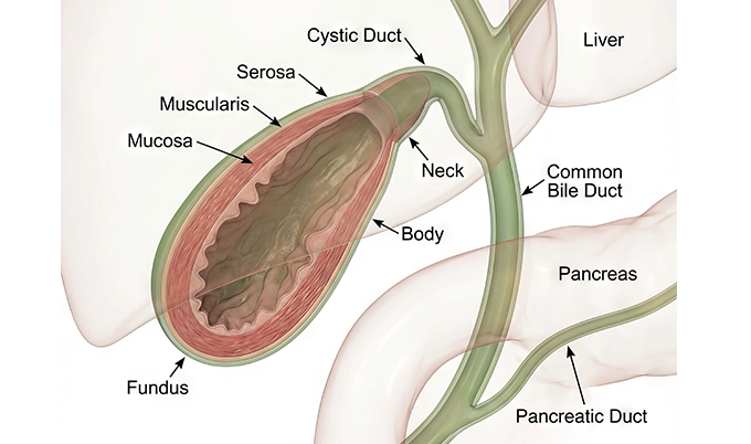
The pain arrives like a knife under your right ribs – sharp, intense, and impossible to ignore. You just finished dinner, maybe something rich or fatty, and now this. Is it indigestion? A heart problem? Or could it be your gallbladder?
Gallstones affect 10-15% of adults in the United States, with most people never knowing they have them. But when stones cause symptoms, the pain can send you to the emergency room, wondering what’s happening inside your body. Understanding the warning signs and how doctors use gallstone imaging to diagnose disease and recognize when that pain demands medical attention.
Recognizing Symptoms With Gallstone Imaging: When Your Body Sends Warning Signals
Most gallstones are “silent” – they sit in your gallbladder causing no problems whatsoever. You could have multiple stones for years without ever feeling them. Then one day, a stone moves into the wrong position, blocks a bile duct, and suddenly your body makes it very clear something is wrong.
The hallmark symptom is biliary colic, which is medical terminology for the specific type of pain gallstones create. This isn’t ordinary stomach discomfort. Biliary colic produces steady, intense pain in your upper right abdomen that builds to a peak and holds there. The pain typically lasts 30 minutes to several hours, though it can persist longer. Unlike the cramping waves you’d feel with intestinal issues, gallstone pain remains constant throughout an episode.
Location matters when identifying gallstone pain. Most people feel it in the upper right quadrant of the abdomen, directly under the right ribcage where your gallbladder sits. But pain doesn’t always stay put. It frequently radiates to your back, particularly between your shoulder blades, or to your right shoulder. Some people feel gallstone pain in the middle of their chest or upper abdomen, which can mimic heartburn or even feel like a heart attack.
Timing provides another critical clue. Gallstone attacks typically strike after meals, especially heavy or fatty ones. When you eat, your gallbladder contracts to release bile that helps digest fats. If a stone is partially blocking a duct, that contraction creates pressure behind the obstruction – and that’s when pain hits. Many gallstone attacks occur in the evening or during the night, hours after dinner.
Beyond pain, watch for these accompanying symptoms. Nausea and vomiting frequently occur during gallstone attacks. Some people develop indigestion, bloating, or excessive belching. If you notice clay-colored stools or dark urine, this suggests bile isn’t flowing properly. Jaundice – yellowing of your skin and eyes – signals that bile is backing up into your bloodstream rather than draining into your intestines.
When Symptoms Signal Something Serious
Biliary colic represents a warning. The pain eventually stops when the stone shifts position and bile can flow again, but this isn’t the end of the problem. Once you’ve had one gallstone attack, you’re likely to have more. And each blockage raises the risk that a stone will stay lodged, triggering serious complications.
Acute cholecystitis develops when a stone completely blocks your gallbladder’s outlet for more than a few hours. The trapped bile causes inflammation and potentially infection. Pain becomes continuous rather than episodic, and you’ll develop fever and chills. The gallbladder wall may become inflamed and swollen. Without treatment, the gallbladder tissue can die, leading to rupture and life-threatening infection in your abdomen.
Cholangitis occurs when stones block the common bile duct, causing infection in your bile ducts. This produces severe right upper abdominal pain, high fever, chills, and jaundice. Cholangitis can progress to sepsis – a body-wide infection that’s a medical emergency. If you have these symptoms, you need immediate care.
Gallstone pancreatitis happens when a stone blocks the pancreatic duct or the shared opening where both the bile duct and pancreatic duct drain into your small intestine. The trapped pancreatic enzymes start digesting the pancreas itself, causing severe inflammation. Pain is intense and often felt across the upper abdomen or boring through to the back. Nausea and vomiting are severe. Gallstone pancreatitis causes 35-40% of all acute pancreatitis cases and can become life-threatening.
Ultrasound: The Gold Standard for Gallstone Detection
When doctors suspect gallstones, ultrasound is almost always the first imaging test they order. For detecting gallstones specifically, ultrasound achieves sensitivity of 88-94% and specificity of 91-95%, making it the most reliable non-invasive method. It’s also quick, painless, involves no radiation, and costs significantly less than CT scanning.
The procedure takes 15-30 minutes and happens while you’re awake. A technologist applies warm gel to your abdomen and moves a handheld transducer across your skin, particularly the upper right area where your gallbladder sits. The device sends sound waves into your body and creates images from the echoes that bounce back. You might need to hold your breath briefly for certain views, and the technologist may ask you to change positions to see the gallbladder from different angles.
On ultrasound, gallstones have a characteristic appearance that makes them easy to identify. They show up as bright white (hyperechoic) spots inside the dark fluid-filled gallbladder. Behind each stone, you’ll see a dark shadow called acoustic shadowing – this happens because ultrasound waves can’t penetrate through the stone, creating a shadowlike area behind it. This shadowing appears regardless of what the stone is made of, whether it’s cholesterol, calcium, or bilirubin.
One unique advantage of ultrasound is the ability to demonstrate stone mobility. The technologist can watch stones move in real-time as you change position. If you roll onto your side, gravity-dependent stones will shift to the lowest part of the gallbladder. This mobility helps distinguish gallstones from gallbladder polyps, which stay attached to the gallbladder wall and don’t move around.
Ultrasound excels at showing complications beyond just finding stones. Gallbladder wall thickening suggests inflammation from cholecystitis. Fluid around the gallbladder indicates infection or inflammation. A distended, enlarged gallbladder may signal obstruction. The technologist can check for a “sonographic Murphy’s sign” by pressing the transducer directly over your gallbladder – if this causes severe pain, it’s a strong indicator of acute cholecystitis. Ultrasound can also evaluate your bile ducts for signs of blockage or dilation.
Limitations exist despite ultrasound’s excellent accuracy. Body habitus affects image quality – excess abdominal fat makes it harder for sound waves to penetrate to the gallbladder. Bowel gas can obscure the view, since ultrasound waves don’t travel well through air. A gallbladder that’s contracted and empty can be difficult to visualize. Very small stones under 2-3mm may be missed. And ultrasound struggles to see the entire length of the bile ducts, particularly the lower portion where it passes behind other organs.
You may need to fast before a gallbladder ultrasound – typically 4-6 hours without eating. Fasting allows your gallbladder to fill with bile, making it larger and easier to visualize. A full gallbladder also makes any stones inside more obvious. Some facilities schedule gallbladder ultrasounds first thing in the morning so you can fast overnight.
Results are typically available within a few hours to a day, though emergency situations may get faster interpretation. The radiologist reviews the images and sends a report to your doctor detailing whether gallstones are present, how many, their size, and any signs of complications. At Craft Body Scan, ultrasound imaging provides this same detailed gallbladder evaluation with transparent pricing and quick appointment availability.
CT Scanning: When Ultrasound Isn’t Enough
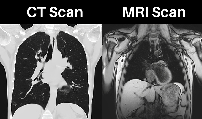
CT scans aren’t the first choice for diagnosing simple gallstones, but they play an important role in specific situations. The sensitivity of CT for detecting gallstones sits around 75% – lower than ultrasound – because many stones don’t show up clearly on CT images. But CT excels at evaluating complications from gallstone disease and identifying other conditions that might be causing your symptoms.
The detection rate depends entirely on stone composition. Only 15-20% of gallstones contain enough calcium to be “radiopaque” – visible on CT scans. These calcified stones appear bright white and are easy to spot. The remaining 80-85% of gallstones are made primarily of cholesterol or bilirubin. On CT, cholesterol stones may appear darker than the surrounding bile (hypoattenuating) or exactly the same density as bile (isodense). When stones match bile density, they’re essentially invisible on CT.
Doctors order CT scans instead of or in addition to ultrasound in several scenarios. If you’re in the emergency department with severe abdominal pain and the cause isn’t clear, CT often becomes the first test because it can identify numerous conditions beyond gallstones – appendicitis, diverticulitis, bowel obstruction, kidney stones, aortic aneurysm, and more. CT provides a comprehensive view of your entire abdomen and pelvis in one scan.
When acute cholecystitis is suspected, CT shows complications that ultrasound might miss. A thickened, inflamed gallbladder wall appears clearly. Pericholecystic fluid and fat stranding around the gallbladder indicate inflammation. Gas within the gallbladder wall suggests gangrenous cholecystitis, a severe complication requiring emergency surgery. Abscesses, gallbladder rupture, and perforation all show up distinctly on CT.
For evaluating gallstone pancreatitis, CT is invaluable. The scan shows pancreatic enlargement and inflammation. Collections of fluid around the pancreas indicate severe disease. CT can identify other causes of pancreatitis and assess the extent of inflammation throughout the abdomen. While ultrasound can diagnose gallstones, CT provides better visualization of the pancreas itself.
CT also helps when ultrasound findings are equivocal or inconclusive. If ultrasound shows a filling defect in the gallbladder but can’t determine whether it’s a stone, polyp, or tumor, CT can provide additional information. When symptoms suggest gallbladder disease but ultrasound appears normal, CT might catch stones that ultrasound missed or identify an entirely different problem causing similar symptoms.
The scan itself takes just 5-10 minutes. You lie on a table that slides into a large donut-shaped machine. The CT scanner rotates around you, taking multiple X-ray images from different angles. A computer processes these images into detailed cross-sectional views. For gallbladder evaluation, contrast dye is often given through an IV to help visualize blood flow and inflammation. The contrast makes inflamed tissues light up, clearly showing areas affected by infection or inflammation.
Radiation exposure is the main drawback. A single abdominal and pelvic CT delivers approximately 10 mSv of radiation – roughly equivalent to three years of natural background radiation. This level is generally safe for occasional use, but repeated CT scans add up. For patients with recurrent gallstone issues requiring multiple imaging studies, minimizing radiation becomes important.
Other Imaging Methods for Specialized Situations
Beyond ultrasound and CT, specialized imaging techniques address specific diagnostic challenges. MRCP (Magnetic Resonance Cholangiopancreatography) provides radiation-free, detailed visualization of bile ducts – particularly valuable for suspected duct stones that ultrasound can’t see. HIDA scans use radioactive tracers to evaluate gallbladder function when symptoms suggest disease but imaging shows no stones. Endoscopic ultrasound combines endoscopy with ultrasound for close-up views that can catch tiny bile duct stones. ERCP serves double duty as both diagnostic and therapeutic, allowing doctors to remove bile duct stones during the same procedure that identifies them.
How Doctors Choose the Right Imaging Test
For straightforward suspected gallstones with classic biliary colic symptoms, ultrasound is the clear first choice. The American College of Radiology recommends ultrasound as the initial test for right upper quadrant pain. Studies show ultrasound detects 88-94% of gallstones, making it the most reliable initial imaging option.
Emergency departments often default to CT for practical reasons – it’s available 24/7, while ultrasound technologists may not be on-site during evening shifts. One study found 52% of gallbladder CT scans occurred between 7pm and 7am when ultrasound was unavailable. However, in 34% of cases reviewed, ultrasound would have been more appropriate based on symptoms.
CT becomes necessary when complications are suspected – acute cholecystitis, cholangitis, pancreatitis, or perforation. The scan identifies inflammation, infection, and involvement of surrounding organs, providing the comprehensive view emergency situations demand.
Pregnancy and younger age favor ultrasound to avoid radiation. Cost also matters – ultrasound runs $200-500 versus $500-3,000 for CT. Starting with the less expensive, more accurate test makes medical and financial sense when symptoms point clearly to gallbladder disease.
What Happens After Imaging Confirms Gallstones

Silent gallstones discovered incidentally rarely need treatment – about 75% of people with gallstones never develop symptoms. Watchful waiting is appropriate for asymptomatic stones.
Once you’ve had biliary colic, surgical removal of the gallbladder (cholecystectomy) becomes the standard recommendation. About 25% of people who have one attack develop serious complications within 10-20 years without treatment. Laparoscopic surgery through small incisions is typical, with most patients going home the same day or after one overnight stay. Recovery takes 1-2 weeks, and the gallbladder isn’t essential for normal digestion.
When surgery isn’t possible, alternatives include medications that dissolve cholesterol stones (though this takes months and stones often recur) or shock wave lithotripsy to break stones into passable pieces. For bile duct stones, ERCP can remove them endoscopically, though the gallbladder typically still requires eventual removal.
Recognizing When Symptoms Need Emergency Care
Certain symptoms signal complications that demand immediate medical attention rather than waiting for an appointment with your doctor.
Seek emergency care if you develop severe, persistent abdominal pain lasting more than a few hours – especially if it’s not resolving like previous biliary colic episodes. Fever combined with right upper abdominal pain suggests infection in the gallbladder or bile ducts. Jaundice indicates bile duct blockage and requires urgent evaluation. Severe nausea and vomiting that prevents you from keeping down liquids can lead to dehydration. Chest pain, especially in older adults or those with heart disease risk factors, should always be evaluated immediately since gallstone pain can mimic heart attacks.
These warning signs indicate that a gallstone has caused a complete blockage or triggered infection. Both situations can deteriorate rapidly without treatment. Emergency departments have the imaging capabilities and surgical backup to diagnose and manage these complications 24/7.
Taking Control of Your Gallbladder Health
Understanding gallstone symptoms and the role of imaging helps you recognize when that upper abdominal pain requires medical evaluation. Biliary colic serves as an early warning that gallstones are causing problems, even though the pain eventually subsides. Ignoring these warning episodes risks serious complications down the line.
Ultrasound remains the gold standard for gallstone diagnosis – over 95% sensitive, radiation-free, and readily available. CT scanning provides essential information when complications are suspected or when ultrasound results are unclear. Together, these imaging methods give doctors the detailed information needed to distinguish simple gallstones from dangerous complications.
If you’re experiencing episodes of upper abdominal pain after meals, especially if it radiates to your back or shoulder, don’t wait for the pain to become severe. Early diagnosis through appropriate imaging allows you to make informed decisions about treatment before emergency complications develop.
At Craft Body Scan, advanced imaging services provide the diagnostic clarity you need to understand what’s happening in your body. When symptoms raise concerns about your gallbladder or other abdominal organs, transparent pricing and efficient scheduling eliminate barriers to getting answers. Schedule your imaging evaluation and take the first step toward resolving your symptoms.