
The pain hits without warning – a sharp, stabbing sensation in your back that makes you double over. If you’ve experienced kidney stone pain, you know there’s nothing quite like it. When that pain strikes, the last thing you want is confusion about which test will give you answers fastest.
In emergency rooms and imaging centers across the country, CT scans detect kidney stones in under 5 minutes – making them the gold standard for rapid diagnosis. But the choice between different imaging methods isn’t always straightforward, and understanding why doctors select specific scans can help you advocate for faster, more accurate care when you need it most.
Why Speed Matters in Kidney Stone Detection

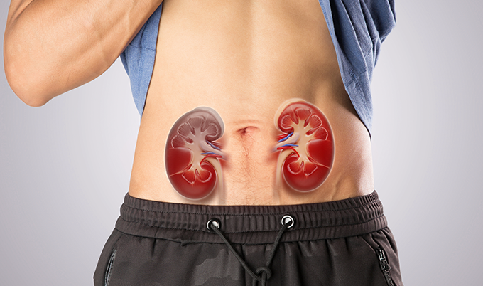
When a kidney stone blocks your urinary tract, every minute counts. The obstruction creates pressure that can damage your kidney if left untreated, and the pain can be absolutely debilitating. I’ve worked with countless patients who’ve described kidney stone pain as worse than childbirth – and they needed answers immediately, not hours later.
Quick diagnosis matters for three critical reasons. First, rapid imaging confirms whether you actually have a kidney stone or if your symptoms point to something else entirely – appendicitis, ovarian cysts, or other conditions that mimic stone pain. Second, the scan shows exactly where the stone is located and how large it is, which determines your treatment path. Third, imaging reveals whether the stone is causing complications like infection or severe blockage that require emergency intervention.
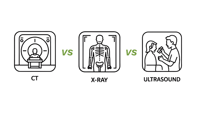
Different imaging methods offer different speeds, accuracy levels, and information quality. The fastest option isn’t always the best option, and the most detailed scan isn’t always necessary. Your doctor weighs multiple factors when choosing which imaging method will serve you best.
CT Scan: The Fastest and Most Accurate Option
Non-contrast CT scanning has revolutionized kidney stone diagnosis. The scan takes 5 to 10 minutes from start to finish, and radiologists can identify stones as small as 1-2 millimeters – about the size of a grain of sand. With sensitivity of 95-96% and specificity of 96-100%, this speed and precision make CT the preferred imaging method in most emergency situations.
The technology works by taking hundreds of cross-sectional images of your abdomen and pelvis. Unlike X-rays, which show a single flat image, CT creates a three-dimensional map that reveals stones regardless of their composition. Calcium stones, uric acid stones, struvite stones – CT catches them all because it’s detecting density differences rather than relying on the stone’s ability to absorb X-rays.
Here’s what makes CT particularly valuable: the scan doesn’t just find the stone. It measures the exact size, shows the precise location in your urinary tract, and reveals whether the stone is causing swelling in your kidney (hydronephrosis). This comprehensive information allows your doctor to predict whether you’ll likely pass the stone on your own or need intervention.
The standard protocol uses no contrast dye, which means no IV, no injection, and no allergic reaction risk. You lie on the table for a few minutes, the machine whirs around you, and you’re done. Results are typically available within 30 minutes to an hour, depending on your facility’s radiology workflow.
Most emergency departments and imaging centers charge between $500 and $3,000 for a kidney stone CT scan, with the average around $1,200 without insurance. At Craft Body Scan, abdominal CT imaging provides this same rapid, detailed visualization at transparent pricing – giving you access to the diagnostic tool doctors trust most for kidney stone detection.
Ultrasound: The Radiation-Free Alternative
Ultrasound imaging offers a completely different approach to finding kidney stones. Instead of radiation, it uses sound waves to create images of your kidneys and urinary tract. The scan takes 20 to 30 minutes, making it slower than CT but still reasonably quick for diagnostic imaging.
Doctors typically choose ultrasound in three specific situations. Pregnant women need stone diagnosis without radiation exposure to the developing baby. Children benefit from avoiding radiation whenever possible, since they’re more sensitive to its effects and have more years ahead for any potential long-term consequences. Patients who require multiple imaging studies throughout their lives – like those with recurrent stones – accumulate less radiation by using ultrasound when appropriate.
The technology has clear strengths and notable limitations. Ultrasound excels at showing whether your kidney is swollen from obstruction, which indicates a stone is blocking flow somewhere in your urinary system. It’s also quite good at finding stones that are sitting in your kidney or at the junction where your ureter meets your bladder. Cost runs significantly lower than CT – typically $200 to $500.
But here’s where ultrasound falls short: it struggles with stones in the ureter, particularly the mid-ureter region, missing them 50-70% of the time. The ureter runs behind your intestines and other structures, and ultrasound waves can’t always penetrate through all that tissue to get a clear view. For renal stones sitting in the kidney itself, ultrasound performs better – with sensitivity around 54-81% depending on stone size and patient body type. Small stones under 3mm often don’t show up clearly on ultrasound. And the test depends heavily on the technician’s skill – an experienced ultrasonographer will find more stones than someone less practiced.
When I talk with patients about ultrasound, I explain it this way: if your ultrasound shows a stone, that’s definitive. But if it doesn’t show a stone and you’re still having symptoms, you may need a CT scan to be certain. Ultrasound is excellent for confirming stones in certain locations and checking for complications, but it’s not the most sensitive tool for initially diagnosing kidney stones.
X-Ray: The Traditional Approach with Significant Gaps
Plain abdominal X-rays (KUB – Kidneys, Ureters, Bladder) were once the standard for kidney stone imaging. The scan takes just a few minutes and costs $100 to $300, making it the fastest and most affordable option. But speed and cost don’t tell the whole story.
X-rays work by detecting calcium, which is why they’re great for finding bones and calcium-based kidney stones. About 80-90% of kidney stones contain calcium, so theoretically, X-rays should catch most stones. In practice, the detection rate ranges from 50-67% for stones, with significant variability depending on stone location and patient factors. That means X-rays miss a substantial portion of stones that are actually present.
The problem comes down to overlapping structures. Your intestines, bones, and other soft tissues create shadows and patterns that can hide stones or create false appearances of stones that aren’t really there. Small stones simply don’t show up. And the 10-20% of stones made from uric acid or certain other materials are completely invisible on X-ray because they don’t contain calcium.
Most modern emergency departments have moved away from using X-rays as the primary diagnostic tool for suspected kidney stones. The technology still has a role – tracking a known stone’s position over time, checking whether a stone has passed after treatment, or providing a quick look when resources are extremely limited. But for initial diagnosis when you’re in acute pain and need definitive answers, X-ray simply doesn’t provide the accuracy that CT offers.
How Doctors Choose Between Imaging Methods
The decision tree that physicians follow isn’t random. Several key factors guide which scan makes the most sense for each patient’s situation.
Clinical urgency plays a major role. If you’re in the emergency department with severe pain, fever, and vomiting – signs that suggest a stone with possible infection – you’re getting a CT scan. The situation demands rapid, comprehensive information because infected obstructed kidneys can become life-threatening quickly. When symptoms are less severe or you’re being evaluated in an outpatient setting, doctors have more flexibility to consider alternatives.
Radiation exposure concerns shift the decision toward ultrasound. A single CT scan of your abdomen delivers about 10 mSv (millisieverts) of radiation – roughly equivalent to three years of natural background radiation. That’s generally considered safe for occasional use, but it adds up if you need repeated imaging. Young patients, pregnant women, and people with recurrent stones who’ll need multiple scans over their lifetime benefit from minimizing radiation through strategic use of ultrasound.
Previous imaging history influences the choice. If you’ve had kidney stones before and previous CT scans showed calcium stones, your doctor might opt for X-ray or ultrasound to track the current stone’s progress. Known stone composition and typical stone patterns in your medical history provide context that makes less sensitive imaging methods more useful.
Availability and cost matter more than we’d like to admit. Some facilities don’t have 24-hour CT availability, or the wait time might stretch several hours. Insurance coverage varies, and out-of-pocket costs can influence which test patients agree to undergo. In settings where CT isn’t immediately accessible, ultrasound provides valuable information while you’re waiting or serves as the definitive test if the clinical picture is clear enough.
Body habitus affects image quality. Ultrasound works less effectively in patients with significant abdominal obesity because sound waves don’t penetrate as well through adipose tissue. CT maintains consistent image quality regardless of body size, though larger patients receive slightly higher radiation doses.
Special Imaging Protocols for Complex Cases
Standard protocols work well for typical kidney stone cases, but certain situations call for modified approaches. Understanding these variations helps you know what to expect if your case requires something beyond routine imaging.
Low-dose CT protocols have emerged as a middle ground between standard CT and other imaging methods. These scans reduce radiation by 40-60% compared to conventional CT while maintaining good image quality for stone detection. The catch? Low-dose protocols work best for average-sized patients and may miss very small stones or subtle findings that standard-dose CT would catch. Many emergency departments now use low-dose protocols as their first-line approach for suspected kidney stones in appropriate patients.
Dual-energy CT represents cutting-edge technology that can actually differentiate stone types. Traditional CT shows you where the stone is and how large it is, but can’t tell you whether it’s a calcium stone, uric acid stone, or something else. Dual-energy CT uses two different X-ray energies simultaneously to analyze the stone’s composition. This information guides treatment – uric acid stones can sometimes be dissolved with medication, while calcium stones typically require different approaches. The technology isn’t yet widely available, and it costs more than standard CT, but it’s becoming increasingly common at larger medical centers.
MRI rarely gets used for kidney stone diagnosis because it doesn’t visualize stones directly. MRI shows soft tissues beautifully but struggles with the calcium and mineral deposits that make up kidney stones. However, MRI can demonstrate the secondary effects of stones – the swelling in your kidney, fluid collections, or complications. In pregnant patients who need more information than ultrasound provides but shouldn’t receive CT radiation, MRI sometimes fills the gap.
What to Expect During Your Kidney Stone Imaging
Knowing what happens during each type of scan reduces anxiety and helps you prepare. Here’s the practical reality of undergoing kidney stone imaging.
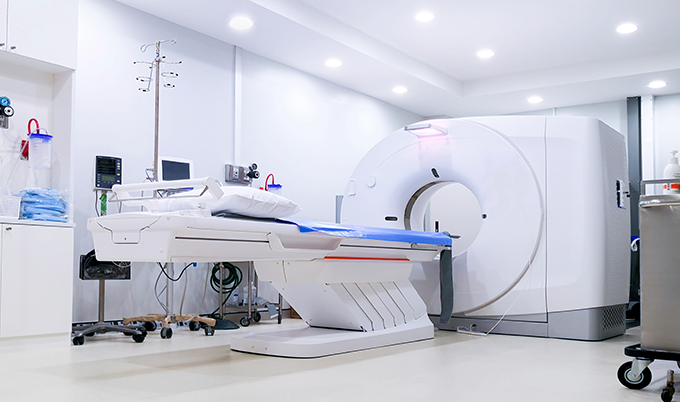
For CT scanning, you’ll change into a gown and remove any metal objects. The technologist positions you on a narrow table that slides into a large donut-shaped machine. You’ll need to lie still and hold your breath for 10-20 seconds while the scanner acquires images. The machine makes whirring and clicking sounds, but the process is painless. No claustrophobia issues – your head stays outside the scanner and the opening is wide. Total time in the room runs 5-10 minutes.
Ultrasound requires a full bladder for optimal kidney imaging, so you may be asked to drink water and wait before your scan. You’ll lie on an exam table while the technologist applies warm gel to your abdomen and back. The ultrasound probe moves over your skin, and you might feel some pressure as the technologist presses to get better images. The process takes 20-30 minutes and involves no radiation or discomfort beyond the pressure of the probe.
X-ray happens fastest – you stand or lie down, hold still for a few seconds while the X-ray is taken, and you’re done. Multiple views may be needed, but each exposure takes only seconds. Total time runs 5 minutes or less.
Results timing varies by setting. Emergency departments typically have radiologists available to read urgent CT scans within 30-60 minutes. Outpatient imaging centers may take several hours to a full day for official results, though critical findings get communicated immediately. Ultrasound and X-ray follow similar timeframes.
Beyond Initial Diagnosis: Follow-Up Imaging
Finding the stone is just the beginning. Follow-up imaging helps track whether conservative management is working or if you need intervention.
Small stones under 5mm pass spontaneously about 65-68% of the time, usually within days to weeks. Your doctor might recommend repeat imaging in 1-2 weeks if you’re managing symptoms at home and trying to pass the stone naturally. This follow-up usually involves X-ray or ultrasound rather than another CT scan – there’s no need for radiation exposure when you’re just checking whether the stone has moved or passed.
Larger stones or stones that haven’t moved after several weeks trigger discussions about surgical intervention. Before procedures like ureteroscopy or shock wave lithotripsy, your surgeon needs current imaging to plan the approach. After procedures, follow-up imaging confirms all stone fragments have been removed or passed.
Patients with recurrent kidney stones need long-term imaging strategies that balance diagnostic accuracy with radiation safety. Some stone formers will have 5, 10, or 20 stones throughout their lifetime. Smart imaging protocols rotate between different methods, use low-dose CT when possible, and avoid unnecessary scans when clinical symptoms clearly indicate what’s happening.
Making Informed Decisions About Your Stone Imaging
When you’re in pain and need answers, you’re not always in the best position to question your doctor’s imaging recommendations. But understanding the options empowers you to have meaningful conversations about your care.
Ask specific questions: “Why are we choosing this imaging method?” “What are we looking for beyond just finding the stone?” “If this scan doesn’t show a stone but I’m still having symptoms, what’s the next step?” These questions help you understand the diagnostic strategy and what to expect.
If radiation exposure concerns you, discuss it directly. Maybe you’ve had multiple CTs in the past year for various medical issues. Maybe you’re planning to start a family soon. Your doctor can’t address concerns you don’t voice, and legitimate reasons for minimizing radiation often exist. The conversation might lead to ultrasound as a first step, or it might clarify why CT is truly necessary in your situation.
Cost transparency matters. Before undergoing imaging, ask about the total cost including the facility fee, radiologist reading fee, and any other charges. Facility prices vary dramatically – the same CT scan might cost $600 at one center and $2,500 at another. Craft Body Scan provides transparent pricing for abdominal imaging, allowing you to plan financially for diagnostic testing without surprise bills weeks later.
Second opinions on imaging interpretation are reasonable when treatment decisions hinge on the results. Radiologist readings are generally accurate, but subtle findings or borderline cases sometimes benefit from review by another specialist. If your doctor recommends surgery based on imaging findings, asking for the images to be reviewed by your surgeon’s radiology colleagues is standard practice, not an insult to your initial radiologist.
The Bottom Line on Kidney Stone Imaging Speed
CT scanning detects kidney stones fastest – 5 to 10 minutes from scan start to completion, with results typically available within an hour. The technology offers unmatched accuracy, catching stones as small as 1-2mm regardless of composition. For patients in acute pain who need immediate answers, CT remains the gold standard.
But “fastest” doesn’t always mean “best” for every situation. Ultrasound provides radiation-free imaging in 20-30 minutes, making it ideal for pregnant women, children, and patients with recurrent stones. X-ray takes just minutes but misses up to half of all stones, limiting its usefulness for initial diagnosis. Your doctor weighs clinical urgency, radiation concerns, previous imaging history, cost, and availability when selecting the most appropriate test for your specific case.
The most important factor isn’t which scan your doctor orders – it’s getting timely imaging that provides the information needed to guide your treatment. Delayed diagnosis leads to prolonged pain, potential complications, and unnecessary suffering. Whether through CT, ultrasound, or X-ray, prompt imaging when kidney stone symptoms strike can make the difference between conservative management and emergency intervention.
At Craft Body Scan, advanced CT imaging technology provides the rapid, accurate diagnosis that kidney stone cases demand. When you need clear answers about abdominal pain or suspected kidney stones, transparent pricing and quick appointment availability eliminate barriers to getting the imaging you need. Take control of your diagnostic journey – schedule your scan and get the clarity you deserve.