After a head injury, doctors need answers fast. Is there bleeding inside your skull causing pressure? Are bones fractured and pushing into brain tissue?
Is dangerous swelling building that could cause permanent brain damage? A brain CT head injury scan provides these critical answers within seconds of completing the imaging study.
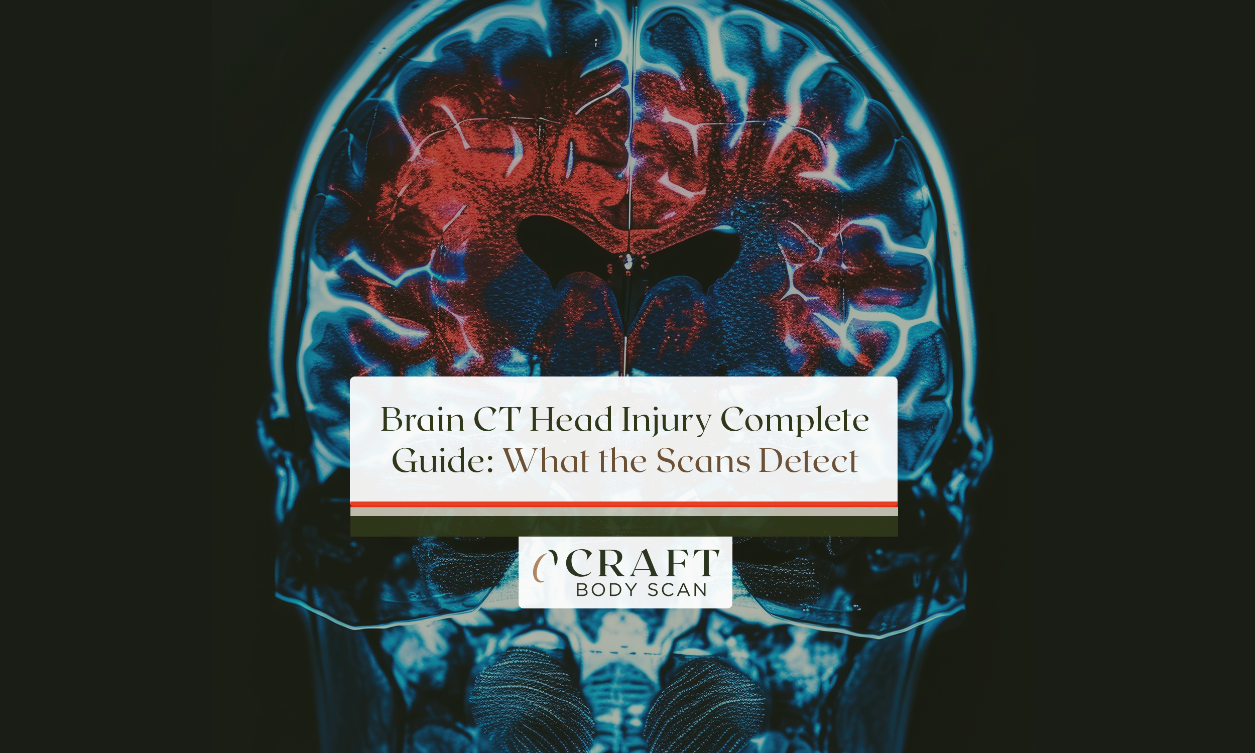
CT (computed tomography) serves as the first-line imaging for traumatic brain injury worldwide. While MRI shows more detail for certain injuries, brain CT head injury scanning offers unique advantages making it the immediate choice in emergency departments.
The scan completes in seconds rather than minutes. Every hospital has CT available 24/7 for emergency CT evaluation.
And the imaging excels at detecting the most dangerous injuries requiring urgent surgical intervention. Understanding what brain CT head injury imaging reveals – and why doctors prioritize it over other tests – helps you make informed decisions after trauma occurs.
The scan detects bleeding, fractures, swelling, and pressure changes visible on CT studies. These findings determine whether you need emergency surgery or can safely go home with observation instructions.
Why Doctors Choose Brain CT Head Injury Imaging First

Emergency physicians face time-critical decisions after head trauma. The primary question: does this patient have a life-threatening injury requiring immediate neurosurgical intervention discovered through brain CT head injury scanning?
Brain CT head injury imaging answers this question faster and more reliably than any other imaging technology currently available. The technology provides immediate information about conditions requiring urgent surgical evacuation.
Speed: Seconds vs Minutes Matter for Brain CT Head Injury
Modern CT scanners complete brain CT head injury imaging in 10-30 seconds total time. The patient lies on the table positioned correctly. The scanner acquires complete images.
You’re done before most people finish reading a single paragraph. This speed advantage proves critical when brain cells are dying or pressure is building inside the skull.
MRI requires 15-30 minutes for equivalent head trauma protocols. Those extra minutes can mean the difference between reversible and permanent brain damage visible on CT studies.
Approximately 1.7 million Americans experience traumatic brain injury annually according to current data. Emergency departments must triage these patients rapidly using CT imaging.
The scan enables fast, accurate decision-making about who needs surgery. It also identifies who can be safely observed without intervention after CT evaluation.
Universal Availability Around the Clock
Every hospital emergency department has CT scanners available 24 hours daily for CT evaluation. Many urgent care facilities now have CT access for after-hours head injury assessment.
This universal availability means brain CT head injury imaging happens immediately when patients arrive. There’s no waiting for specialized equipment to become available during off-hours.
MRI machines are far less available than CT scanners. Smaller hospitals may have only one MRI that requires advance scheduling.
Evening and weekend MRI availability is limited at many facilities. When minutes matter after head trauma, brain CT head injury scanning provides immediate answers without waiting.
Compatibility with Life Support Equipment
Severe head injury patients arrive with multiple monitors, ventilators, and life support devices. These metal-containing devices work normally in CT scanners during CT imaging.
The imaging proceeds without disconnecting critical equipment maintaining the patient alive. MRI’s powerful magnetic field prohibits metal objects entirely from the scanning room.
Ventilators, monitoring equipment, and implanted devices must remain outside the MRI room. This creates logistical nightmares for unstable trauma patients requiring continuous life support during CT assessment.
No Safety Screening Required for Emergency Brain CT Head Injury
CT imaging proceeds immediately without safety screening delays. There are no contraindications preventing emergency brain CT head injury scans in trauma patients who need them urgently.
MRI requires detailed safety screening before scanning begins. Pacemakers, cochlear implants, certain aneurysm clips, and metal fragments contraindicate MRI completely.
After trauma, you may not know what devices or metal fragments are present. Brain CT head injury imaging proceeds without delay while safety questions get sorted out later.
 Why Brain CT Head Injury Is First-Line Imaging
- Speed – Complete scan in 10-30 seconds vs 15-30 minutes for MRI
- Availability – Present 24/7 at all emergency departments
- No contraindications – Works with pacemakers, implants, life support
- Bone detail – Shows skull fractures with exceptional clarity
- Acute hemorrhage – Detects fresh bleeding immediately
- 92% sensitivity – Identifies vast majority of injuries at presentation
- Guides surgery – Shows exact location, size, and urgency of injuries
What Brain CT Head Injury Scans Detect After Trauma

The non-contrast CT provides exquisite detail of bone and acute bleeding. These findings determine immediate treatment decisions after head trauma occurs through CT interpretation.
Skull Fractures and Bone Injuries on Brain CT Head Injury Scans
Brain CT head injury imaging shows skull fractures with remarkable clarity and detail. The scan detects linear fractures, depressed fractures where bone pushes inward, and complex fractures involving multiple bones.
Skull base fractures appear on CT studies when other imaging might miss them. These fractures carry high risk for cerebrospinal fluid leaks and meningitis requiring intervention.
Detecting them early on CT scans enables appropriate antibiotic prophylaxis. It also facilitates neurosurgical consultation for potential repair procedures.
Depressed skull fractures require surgical elevation when bone fragments compress brain tissue. The brain CT head injury scan shows exactly how much bone depression exists underneath.
It reveals whether fragments penetrate the dura covering the brain. This information guides surgical planning for cases requiring intervention.
Epidural Hematoma: The Classic Neurosurgical Emergency
Epidural hematoma forms between skull and dura mater covering the brain. Arterial bleeding creates rapidly expanding blood collections that can kill within hours if untreated.
Brain CT head injury imaging shows these as lens-shaped (biconvex) collections adjacent to skull fractures. The classic epidural hematoma involves the middle meningeal artery running under temporal bone.
The injury occurs in the temporoparietal region 75% of the time. Associated skull fracture is present 90% of the time on CT scans showing this emergency.
Early detection via CT imaging dramatically improves patient outcomes. Surgical evacuation before the patient loses consciousness results in excellent recovery rates.
Delayed diagnosis after herniation develops leads to death or severe disability. This makes rapid brain CT head injury scanning critical for survival.
Subdural Hematoma: Blood Between Brain and Dura
Subdural hematomas collect between dura and brain surface creating pressure. Venous bleeding creates crescent-shaped collections that may extend over large brain areas.
Brain CT head injury scans show these as concave collections following brain contours. Acute subdural hematomas appear bright white (hyperdense) on CT imaging studies.
They develop immediately after trauma from torn bridging veins. Large subdurals causing mass effect require urgent surgical drainage guided by brain CT head injury findings.
Chronic subdural hematomas develop days to weeks after initial injury. They appear dark (hypodense) on CT as blood breaks down over time.
Elderly patients and those on blood thinners are particularly vulnerable. Serial brain CT head injury scans track enlargement requiring surgical intervention.
Subarachnoid Hemorrhage: Bleeding in CSF Spaces
Traumatic subarachnoid hemorrhage appears in CSF-filled spaces around the brain. Blood layers along brain surface and fills sulci between gyri.
Brain CT head injury imaging shows this as increased density outlining brain contours. Basilar cistern hemorrhage indicates severe trauma requiring intensive monitoring.
This blood pattern suggests rotational forces or direct basilar skull impact. Isolated basilar hemorrhage on CT scans may indicate underlying vascular injury.
Such patterns require angiography to rule out traumatic aneurysm. The brain CT head injury findings guide additional imaging decisions.
Intracerebral Hemorrhage and Brain Contusions
Intracerebral hemorrhage involves bleeding directly into brain tissue parenchyma. These appear as bright white areas within brain substance on CT studies.
Location determines symptoms visible clinically – frontal lobe hemorrhage affects personality and judgment. Occipital lobe bleeding causes vision loss requiring different management approaches.
Cerebral contusions are brain bruises visible on brain CT head injury imaging. They appear as mixed density areas combining bleeding and edema swelling.
Contusions commonly occur at frontal and temporal poles. Brain impacts rough skull surfaces during trauma causing brain CT head injury-visible damage.
Contusions evolve over time requiring follow-up imaging. Approximately 25-45% increase in size within the first 24 hours after initial trauma.
This progression explains why repeat brain CT head injury imaging is often performed 6-12 hours later. The follow-up scan captures evolving injuries missed on initial studies.
| Injury Type | CT Appearance | Clinical Significance | Urgency Level |
|---|---|---|---|
| Epidural Hematoma | Lens-shaped, biconvex | Arterial bleeding, rapid expansion | EMERGENCY – hours matter |
| Subdural Hematoma | Crescent-shaped, follows contours | Venous bleeding, variable speed | URGENT – depends on size |
| Subarachnoid Hemorrhage | Blood in CSF spaces, sulci | May indicate vascular injury | URGENT – requires monitoring |
| Cerebral Contusion | Mixed density, brain bruising | 25-45% expand within 24 hours | MODERATE – needs follow-up |
| Skull Fracture | Bone discontinuity, fragments | 90% associated with epidural | VARIABLE – depends on type |
Diffuse Axonal Injury: Brain CT Head Injury Limitations
Diffuse axonal injury involves tearing of nerve fibers throughout the brain. This injury pattern doesn’t show well on CT despite causing severe symptoms requiring intensive care.
Small hemorrhages at gray-white matter junctions may be the only visible sign on CT scans. Most axonal damage remains invisible on conventional CT imaging studies.
MRI with specialized sequences detects diffuse axonal injury far better than CT. When symptoms seem severe but brain CT head injury imaging appears relatively normal, MRI often reveals extensive damage.
The MRI findings explain the clinical picture invisible on CT studies. This represents one of CT’s main limitations for traumatic brain injury evaluation.
Cerebral Edema and Dangerous Brain Swelling
Brain swelling develops hours after trauma as inflammatory response begins. CT shows loss of normal gray-white matter differentiation as brain swells against skull.
Sulci disappear and ventricles shrink as edema increases on serial CT studies. Severe edema causes herniation syndromes requiring emergency intervention.
Brain tissue shifts across normal boundaries compressing vital structures. Uncal herniation compresses the midbrain controlling consciousness and breathing.
Subfalcine herniation pushes structures across midline creating additional damage. These herniation patterns visible on CT scans indicate life-threatening pressure.
Immediate intervention becomes necessary to prevent death. The brain CT head injury findings guide decompressive surgery decisions.
Clinical Decision Rules for Brain CT Head Injury Imaging
Not every head bump requires head trauma CT exposure to radiation. Radiation exposure and healthcare costs demand selective imaging based on clinical criteria.
Two validated clinical decision tools guide brain CT head injury ordering decisions. These rules maintain safety while reducing unnecessary scans in low-risk patients.
Canadian CT Head Rule for Brain CT Head Injury
The Canadian CT Head Rule applies to patients with minor head injury (Glasgow Coma Scale 13-15). The rule identifies patients at high and medium risk for neurosurgical intervention based on brain CT head injury findings.
High risk criteria requiring immediate brain CT head injury imaging:
– Glasgow Coma Scale score less than 15 at 2 hours post-injury
– Suspected skull fracture (visible, palpable, bleeding from ears or nose)
– Any sign of basal skull fracture (Battle’s sign, raccoon eyes)
– Two or more vomiting episodes after injury
– Age 65 years or older
Medium risk criteria where brain CT head injury scanning prevents missing important injuries:
– Amnesia before impact exceeding 30 minutes duration
– Dangerous mechanism (pedestrian struck, ejection from vehicle, fall from elevation >3 feet)
This rule demonstrates 100% sensitivity for injuries requiring neurosurgery. When applied correctly to brain CT head injury decisions, no surgical lesions get missed.
New Orleans Criteria for Mild Traumatic Brain Injury
The New Orleans Criteria apply to patients with mild traumatic brain injury and brief loss of consciousness. Any of these findings warrant brain CT head injury imaging without delay:
– Headache complaint after injury
– Vomiting episodes (any number)
– Age over 60 years
– Drug or alcohol intoxication
– Persistent anterograde amnesia (can’t form new memories)
– Visible trauma above the clavicles
– Seizure occurrence after injury
These criteria are highly sensitive for detecting intracranial pathology on CT scans. The trade-off is lower specificity – more scans get performed but fewer injuries get missed entirely.
When Follow-Up Brain CT Head Injury Imaging Is Needed
Initial brain CT head injury scans don’t tell the complete evolving story. Traumatic brain injury is a dynamic process changing over hours and days.
Bleeding can expand significantly. Swelling can progress dangerously. New injuries can develop over hours requiring additional CT evaluation.
Timing of Repeat Brain CT Head Injury Scans
Patients with any abnormality on initial brain CT head injury imaging typically receive repeat scanning 6-12 hours later. This timing captures progression of primary injuries visible initially.
It also identifies development of secondary complications not present earlier. Early scans performed less than 1 hour after injury may miss injuries still developing at cellular levels.
CT sensitivity improves when scanning occurs 1-6 hours after trauma. Very early brain CT head injury imaging sometimes requires repeat scanning even when initial results appear normal initially.
Clinical Deterioration Warrants Immediate Repeat Imaging
Any worsening neurological status demands immediate repeat brain CT head injury scanning regardless of initial findings. New weakness, decreased consciousness, severe headache, or vomiting suggest evolving injury underneath.
Delayed hemorrhage develops in some patients hours after normal initial scans. Anticoagulation and bleeding disorders increase this risk dramatically requiring close monitoring.
Close observation and low threshold for repeat brain CT head injury imaging protects these vulnerable patients. The repeat scan often reveals new bleeding requiring intervention.
 When to Get Repeat Brain CT Head Injury Scan
- Routine timing – 6-12 hours after initial scan showing any abnormality
- Worsening symptoms – New weakness, decreased consciousness, severe headache
- New vomiting – Especially if initial scan was normal
- Seizure activity – Any new seizure after head injury
- Anticoagulation – Patients on blood thinners need close monitoring
- Very early initial scan – If first CT was within 1 hour of injury
- Clinical-radiologic mismatch – Severe symptoms but minimal CT findings
Brain CT Head Injury vs MRI After Trauma

Both imaging modalities have roles in traumatic brain injury evaluation. Understanding when each test adds value helps optimize care after CT assessment.
What MRI Adds Beyond Brain CT Head Injury Imaging
MRI detects diffuse axonal injury invisible on CT scans. The specialized diffusion and susceptibility sequences show microscopic hemorrhages and axonal tears.
These findings explain symptoms despite normal-appearing brain CT head injury scans. Small cortical contusions show better on MRI than on conventional CT imaging.
Posterior fossa injuries in the cerebellum and brainstem appear clearer without CT’s bone artifact. Chronic injuries and their long-term effects become visible on MRI weeks after brain CT head injury imaging showed only acute findings.
When to Add MRI After CT
Consider MRI when symptoms seem severe but brain CT head injury imaging shows minimal abnormalities. Persistent confusion, memory problems, or focal deficits without clear CT explanation warrant MRI investigation thoroughly.
Subacute and chronic evaluation benefits from MRI capabilities. After the acute phase managed with brain CT head injury scans, MRI provides superior detail.
The imaging predicts recovery outcomes better than CT in this time frame. It also guides rehabilitation planning more effectively than CT studies alone.
Why CT Remains First-Line Despite MRI Advantages
Despite MRI advantages for certain injuries, CT maintains its position as initial brain CT head injury imaging modality. The speed, availability, and safety advantages outweigh MRI’s superior soft tissue detail completely.
In acute trauma settings where minutes matter, brain CT head injury scanning provides the information needed immediately. Life-threatening injuries requiring surgery all show well on CT imaging studies.
The injuries that MRI detects better typically don’t require emergency intervention. This makes brain CT head injury scanning the logical first choice for acute trauma evaluation.
What Happens During Emergency Brain CT Head Injury Scanning

Understanding the process reduces anxiety when you or a loved one needs emergency head injury imaging after trauma occurs.
Rapid Triage and Positioning
After emergency department arrival, nurses remove metal objects from your body. They position you on the CT table for brain CT head injury scanning.
A foam holder stabilizes your head preventing movement during imaging. The table slides through the doughnut-shaped scanner opening automatically.
Your head passes through while the scanner rotates around capturing images. The actual image acquisition takes seconds for CT studies to complete.
Image Acquisition and Reconstruction
Modern CT uses thin slices (2.5-5mm) capturing detailed information. Computers reconstruct these slices into images viewed at different “windows” for brain CT head injury interpretation.
Brain window, bone window, subdural window – each optimized for detecting specific findings. Radiologists examine every slice looking for abnormalities visible on brain CT head injury images.
They look for bleeding, fractures, swelling, and mass effect. In trauma cases, interpretation happens immediately without delay.
Results reach the emergency team within minutes of completing brain CT head injury imaging. This speed enables rapid treatment decisions for surgical candidates.
When Contrast Gets Added to Brain CT Head Injury Protocol
Initial brain CT head injury scans use no IV contrast material. Contrast could obscure subtle bleeding requiring detection first.
After confirming no hemorrhage, CT angiography with contrast may follow. This happens when vascular injury is suspected based on fracture patterns visible on initial brain CT head injury scans.
Hemorrhage location may also suggest vascular injury. The brain CT head injury findings guide additional imaging decisions about angiography needs.
Cost and Accessibility of Brain CT Head Injury Imaging
Emergency brain CT head injury scans are considered medically necessary after significant trauma. Insurance typically covers imaging without prior authorization when doctors order it emergently.
Out-of-pocket costs range from $300-$3,000 without insurance depending on facility. Hospital emergency departments charge more than independent imaging centers for CT studies.
Most insurance plans cover emergency brain CT head injury imaging at in-network rates. Federal law requires stabilizing treatment regardless of insurance status or ability to pay.
Never delay seeking care due to cost concerns. Financial counselors can help with billing after your medical emergency is stabilized following CT evaluation.
Long-Term Implications After Brain CT Head Injury Scans
Not all head injuries show immediate abnormalities on CT scans. Understanding when delayed imaging helps guides recovery planning after initial evaluation.
Post-concussion syndrome symptoms can persist months despite normal brain CT head injury imaging. Learn how to check for a concussion to identify symptoms early. The functional changes causing post-concussion syndrome occur at cellular levels invisible on conventional CT.
Advanced imaging techniques including functional MRI may help explain persistent symptoms. But currently, concussion remains largely a clinical diagnosis regardless of brain CT head injury findings.
Repeated head injuries over years may cause chronic traumatic encephalopathy. This degenerative brain disease doesn’t show on routine brain CT head injury imaging protocols.
Specialized imaging research continues investigating ways to detect these changes earlier. Current brain CT head injury protocols focus on acute injuries requiring immediate intervention rather than long-term degeneration.
At Craft Body Scan, we understand the importance of early detection for serious health conditions. While emergency brain CT head injury imaging requires immediate hospital evaluation, our preventive health screening services help identify potential issues before emergencies occur.
Our board-certified radiologists provide detailed interpretations of all imaging studies. According to research from the National Institutes of Health, prompt imaging after head trauma significantly improves outcomes by enabling rapid treatment decisions.
The American College of Radiology provides comprehensive guidelines on appropriate imaging use for head trauma. These evidence-based recommendations help doctors order brain CT head injury scans selectively while maintaining safety standards.
Brain CT head injury imaging serves as the cornerstone of acute head trauma evaluation. The technology’s speed, availability, and accuracy for detecting life-threatening injuries make it irreplaceable in emergency settings.
Understanding what CT shows – and what it misses – helps patients and families navigate the often confusing hours after head trauma. When doctors recommend urgent brain CT head injury scanning, they’re looking for bleeding, fractures, and pressure changes.
These findings determine whether you need surgery or can safely go home. Modern brain CT head injury protocols balance radiation concerns against the critical need for accurate diagnosis determining treatment.
Clinical decision rules help doctors order scans selectively while maintaining safety. The imaging continues evolving with reduced radiation doses and improved detection of subtle injuries visible on CT studies.
If you’ve experienced head trauma, follow your doctor’s recommendations about imaging timing. Initial brain CT head injury scans establish baseline findings for comparison.
Follow-up imaging tracks progression of known injuries. And in some cases, MRI adds information CT cannot provide about recovery and prognosis after CT evaluation.
Schedule your consultation if you have questions about preventive imaging for head injury risk factors. For emergency symptoms after trauma, always seek immediate emergency department evaluation with brain CT head injury scanning as appropriate.