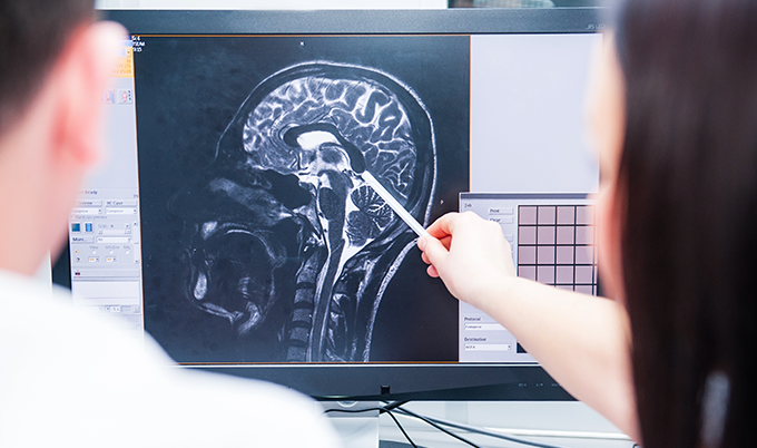
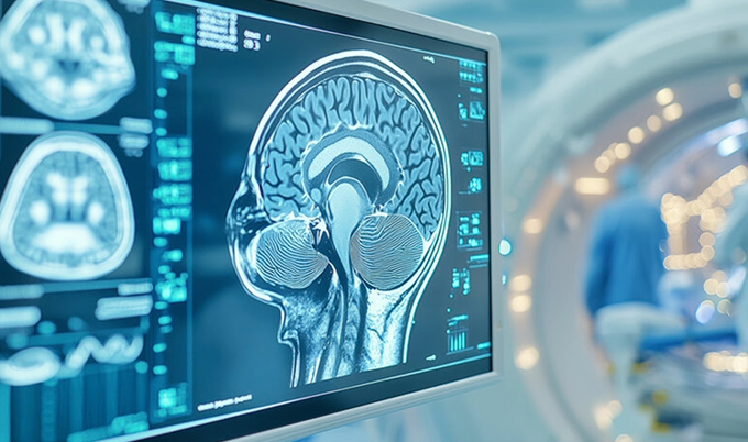
If you’ve just received your brain MRI results, you’re probably feeling a mix of relief and confusion. As radiologists who review hundreds of brain scans each month, we see that same look on patients’ faces when they’re handed their imaging results. The technical language can feel overwhelming. Those black and white images might look foreign and mysterious. You’re left wondering: Is this actually normal?
Here’s what we want you to know: a normal brain MRI shows healthy brain tissue with clear boundaries between gray and white matter, symmetrical structures, and no signs of abnormal growths, inflammation, or damage. But there’s so much more to understanding what “normal” truly means for your specific scan—and why that matters for your peace of mind.
The Anatomy of Normal Brain MRI Results

Think of your brain MRI as creating a detailed photo album of your brain using magnetic fields and radio waves—completely safe, with no radiation involved. It’s like taking thousands of cross-sectional photographs of your brain, with each slice revealing different structures and tissues that tell the story of your brain health.
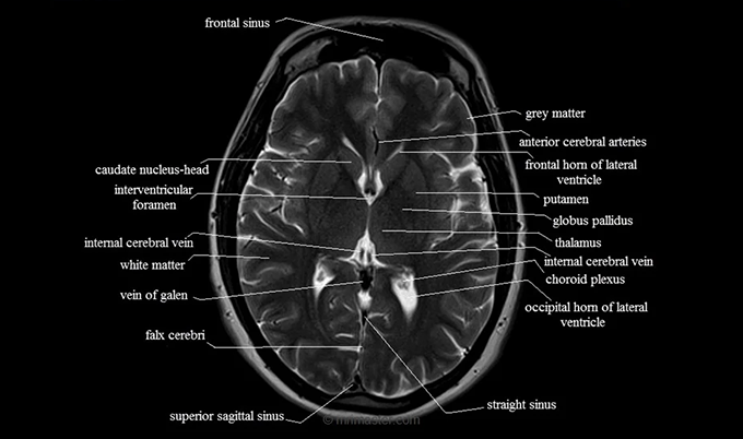
When we review a normal brain MRI, we’re looking for several key features that indicate your brain is functioning well. The cerebral cortex appears as a thin layer of gray matter that wraps around your brain’s surface—think of it as the brain’s outer shell where most of your thinking happens. Beneath this, white matter forms your brain’s communication highways, appearing lighter on most MRI sequences and connecting different brain regions.
Your ventricles—those fluid-filled spaces within your brain—should appear symmetrical and appropriately sized. These cerebrospinal fluid spaces are incredibly important because they help cushion your brain and remove waste products, kind of like your brain’s built-in cleaning system. When they’re too large or irregularly shaped, it might indicate various conditions, but when they look normal, it’s a great sign.
Your brain stem, which connects your brain to your spinal cord, should show clear, well-defined structures. And your cerebellum, the part responsible for balance and coordination, appears at your brain’s base with its characteristic folded appearance—almost like a small, intricate tree.
🧠 Key Features of a Normal Brain MRI
✓ Symmetrical Structure
Left and right brain hemispheres show balanced proportions with no significant shifts or distortions.
✓ Clear Gray/White Matter
Distinct boundaries between gray matter (nerve cells) and white matter (connecting fibers).
✓ Normal Ventricles
Fluid-filled spaces appear appropriately sized and symmetrical throughout the brain.
Understanding these normal features can really help reduce anxiety when you’re reviewing your brain MRI results. What surprises many patients is learning that “normal” doesn’t mean identical to every other brain. Just like fingerprints, each brain has unique characteristics while maintaining essential healthy patterns.
Key Structures Visible in a Normal Brain MRI

Understanding the specific structures we examine as radiologists can help demystify your results. We systematically review each area, looking for signs of health and checking for any deviations from normal patterns. Here’s what we’re seeing when we look at your scan:
Gray Matter Distribution: In a healthy brain, gray matter appears evenly distributed across the cerebral cortex. This tissue contains most of your brain’s nerve cell bodies and appears darker on certain MRI sequences. We measure its thickness and look for symmetry between your left and right hemispheres—it’s like checking that both sides of your brain are working together harmoniously.
White Matter Integrity: White matter consists of nerve fibers that connect different brain regions—imagine them as the brain’s internal internet cables. Normal white matter appears bright on T2-weighted images and shows no areas of abnormal signal intensity. We specifically look for what we call “white matter hyperintensities”—bright spots that can sometimes indicate various conditions.
Ventricular System: Your brain’s ventricles should maintain appropriate size and shape. Normal ventricles appear symmetrical, with the lateral ventricles being the largest. As we age, these can naturally enlarge slightly, and that’s completely normal—it’s just your brain adapting over time.
Blood Vessel Patterns: A normal brain MRI shows clear blood vessel patterns without signs of blockages, aneurysms, or abnormal connections. Your major arteries and veins should follow expected pathways with appropriate size—essentially, your brain’s plumbing should look healthy and well-organized.
Midline Structures: Your brain’s midline structures should remain centered, like a well-balanced house. Any shift could indicate pressure from masses or swelling, so when everything looks centered and in place, that’s exactly what we want to see.
Signal Characteristics: Different tissues produce different signals on MRI, and normal brain tissue shows predictable signal patterns across various imaging sequences. We compare these patterns to identify any abnormalities—think of it as reading your brain’s unique signature.
How Radiologists Interpret Normal Brain Scans

When we review your brain MRI, we follow a systematic approach that we’ve developed over years of training and experience. This methodical process ensures we don’t miss anything important while efficiently providing you with accurate results.
First, we assess image quality. Sometimes motion during the scan, positioning issues, or technical problems can create images that might look concerning but aren’t actually related to your health. Making sure we have a high-quality scan gives us the foundation for accurate interpretation.
Next comes our systematic review. We examine each major brain region, carefully comparing left and right sides for symmetry. Your frontal lobes, responsible for executive function and personality, should show normal gray-white matter differentiation. Your temporal lobes, crucial for memory and language, require careful attention—especially the hippocampal formations that are so important for forming new memories.
Age-related changes present unique interpretation challenges, and this is where our experience really matters. What might appear concerning in a 30-year-old could represent completely normal aging in a 70-year-old. Your brain volume naturally decreases with age, and small white matter changes become increasingly common as part of normal aging.
We pay special attention to areas that tend to show early changes when problems do develop. The periventricular regions, where conditions like multiple sclerosis lesions often first appear, receive careful scrutiny. The basal ganglia, which can be affected by movement disorders, require detailed evaluation.
Each MRI sequence provides different information, like looking at your brain through different filters. T1-weighted images excel at showing anatomy and detecting blood products. T2-weighted images highlight abnormalities and fluid collections. FLAIR sequences suppress cerebrospinal fluid signals, making certain types of lesions more visible.
The final step involves putting together all the imaging findings with your clinical information. A truly normal brain MRI, considered alongside your symptoms and medical history, provides the complete picture we need for optimal patient care.
How Radiologists Review Your Brain MRI
- Image Quality Assessment
We verify scan clarity and check for any technical issues that might affect interpretation. - Systematic Brain Review
Our radiologists examine each brain region systematically, comparing structures for symmetry and normal appearance. - Age-Appropriate Analysis
We consider your age and medical history to distinguish normal aging changes from concerning findings. - Comprehensive Report
We provide clear, detailed results with explanations you can understand and actionable next steps.
Common Variations That Are Still Normal

One of the most important things we want you to understand is that normal brain anatomy includes significant variation between individuals. What might initially look concerning to someone who isn’t trained in reading these scans often represents completely normal anatomical variants.
Asymmetry Within Normal Limits: Perfect symmetry doesn’t exist in human brains, and that’s completely normal. Your right hemisphere is typically slightly larger than your left, and the occipital horns of your lateral ventricles often show size differences. These asymmetries fall within established normal ranges and are nothing to worry about.
Age-Related Changes: As we age, our brains undergo predictable changes that remain within normal parameters. Mild brain volume loss begins in our twenties and accelerates after age 60—this is completely normal and expected. Small white matter hyperintensities become increasingly common and usually represent normal vascular aging, not disease.
Anatomical Variants: Some people have additional sulci (brain folds) or slightly different gyral patterns—think of these as normal variations in your brain’s landscape. Your corpus callosum can show normal thickness variations, and your pineal gland, a small endocrine structure, commonly calcifies with age and appears bright on certain sequences.
Incidental Findings: Sometimes normal brain MRIs reveal incidental findings that require no treatment whatsoever. Small arachnoid cysts, developmental venous anomalies, or tiny areas of gliosis might appear on scans of completely healthy individuals. These are like birthmarks in your brain—present, harmless, and requiring no intervention.
Physiological Changes: Your hydration status, time of day, and recent activities can subtly affect how your brain appears on MRI. Well-hydrated patients might show slightly different ventricular sizes compared to dehydrated individuals—these are normal, temporary variations.
Understanding these variations can prevent unnecessary anxiety when reviewing your results. What matters most is that your brain shows no signs of active disease processes or structural abnormalities requiring intervention.
When to Get a Brain MRI for Preventive Health
Preventive brain imaging can be a powerful tool for early detection of conditions that benefit from prompt intervention. While not everyone needs routine brain MRIs, certain situations make proactive scanning a smart choice for your health.
Family history plays a crucial role in preventive brain imaging decisions. If you have relatives with brain aneurysms, certain types of strokes, or hereditary conditions affecting the brain, early screening can identify risks before symptoms develop. It’s like having a roadmap of potential concerns to watch for.
Persistent neurological symptoms deserve investigation, even when initial evaluations appear normal. Chronic headaches that change in pattern, memory concerns beyond normal aging, or subtle balance issues might warrant brain imaging for peace of mind. Sometimes, having that normal scan result is exactly what you need to feel confident about your health.
Athletes participating in contact sports face unique risks for traumatic brain injury. Baseline brain MRIs can provide valuable comparison points if future head injuries occur. This proactive approach helps optimize treatment decisions and long-term care planning—it’s like having a before photo for comparison.
If you’ve had occupational exposures to certain chemicals, radiation, or other toxins, you might face increased brain disease risks. Individuals with these exposures can benefit from periodic brain imaging to monitor for early changes and catch any problems when they’re most treatable.
Sometimes people simply want the reassurance that comes with knowing their brain health status. Just as we check cholesterol levels and blood pressure regularly, brain imaging can provide valuable baseline information about one of your most critical organs. There’s real value in that peace of mind.
The key is balancing the benefits of early detection against the costs and potential anxiety associated with imaging. A conversation with healthcare providers familiar with your specific situation helps determine the most appropriate screening approach for you.
Next Steps After Receiving Normal Results

Receiving normal brain MRI results should provide genuine reassurance, but it’s also a wonderful opportunity to optimize your ongoing brain health. A normal scan represents a snapshot in time—maintaining that health requires ongoing attention to factors that support cognitive function.
Lifestyle Optimization: Normal brain MRI results give you a strong foundation for implementing brain-healthy lifestyle choices. Regular cardiovascular exercise improves blood flow to your brain and supports the growth of new neural connections. The Mediterranean diet, rich in omega-3 fatty acids and antioxidants, provides nutrients that actively support brain health.
Continued Monitoring: Depending on your risk factors and symptoms, your healthcare provider might recommend follow-up imaging at specific intervals. Some conditions develop slowly over time, making periodic reassessment valuable for early detection. Think of it as regular maintenance for your most important organ.
Symptom Awareness: Having normal baseline results helps you and your healthcare team better evaluate any future neurological symptoms. If new concerns arise, having recent normal imaging provides important context and can actually speed up the diagnostic process.
Documentation and Records: Keep copies of your normal brain MRI results and the radiologist’s report. This documentation becomes incredibly valuable for future healthcare providers and can prevent unnecessary repeat imaging if you move or change doctors.
Preventive Planning: Use your normal results as motivation to maintain habits that support long-term brain health. Regular sleep, stress management, social engagement, and mental stimulation all contribute to preserving cognitive function over time. You’re investing in your future self.
Normal brain MRI results represent more than just the absence of disease—they confirm that your brain shows healthy structure and function. This knowledge empowers you to make informed decisions about your ongoing neurological health and provides genuine peace of mind about one of your body’s most vital organs.
At Craft Body Scan, our board-certified radiologists specialize in providing clear, comprehensive interpretations of brain imaging studies. We understand that medical imaging can feel overwhelming, which is why we focus on explaining results in terms that make sense for your specific situation and help you feel confident about your health.
Ready to take control of your brain health? Schedule your comprehensive brain scan with Craft Body Scan today. Our advanced imaging technology and expert interpretation provide the detailed information you need to make informed decisions about your neurological wellness.
Contact us to learn more about our brain imaging services and how we can support your proactive approach to health.


